Aron Shapiro, Andover, Mass.
No other group of compounds has such a well-documented and potent analgesic effect as opioids. These agents have been used by doctors to control pain since antiquity and are even mentioned in the oldest known medical document, the Ebers Papyrus, which dates back over 3,500 years.1 Unfortunately, opioids have also been abused for millennia and their use in modern medicine is limited due to a broad range of side effects, including nausea, constipation, respiratory depression and addiction.2
Despite their drawbacks, opioids represent a largely untapped resource in ophthalmology. In addition to established evidence of effective ocular anesthesia, preliminary studies suggest that opioids may be viable intraocular pressure-lowering agents and offer a useful alternative to current topical glaucoma therapies.3 Initial research also shows that topically applied opioid receptor antagonists may assist in corneal wound healing and serve as an alternative to current topical ocular anesthetics that delay corneal re-epithelialization.4 While centrally acting opioids such as morphine and codeine cross the blood-brain barrier and have shown a strong potential for abuse, new topical formulations with limited systemic absorption may provide good alternatives to current ocular therapies without the central nervous system side effects of their systemically administered counterparts.5
Pharmacology of Opioids
Modern opioids fit into one of four categories: natural; endogenous; synthetic or semi-synthetic. Natural opioids are alkaloids refined directly from the opium plant, Papaver somniferous. Endogenous opioids, such as endorphin and dynorphin, are produced within humans and act as the body's natural painkillers. Semi-synthetic opioids are derived from natural opium alkaloids, and synthetic opioids are produced in the laboratory without opium extract. All types of opioids produce their clinical effects through the activation (or deactivation) of opioid receptors. This distinct receptor family consists of G-protein-coupled transmembrane proteins found on a variety of cells in the central and peripheral nervous systems.
There are three classical opioid receptors: mu; delta and kappa. While most clinically administered opioids are mu agonists, delta and kappa ligands are also known to inhibit the activity of nociceptors and relieve pain throughout the central and peripheral nervous systems.
Two novel types of opioid receptors, sigma and zeta (also known as opioid growth factor), have also been reported in some research studies. The sigma receptor, however, has since been removed from the opioid receptor family classification because it is activated by non-opioid ligands.6 Opioid growth factor receptors (OGFr), by contrast, share little structural homology with mu, delta and kappa opioid receptors and regulate the cell cycle rather than pain, but are activated by some opioid ligands and are therefore considered to be true opioid receptors.7
Opioid receptors of all types—mu, delta and kappa—have been found on sensory neurons and their expression is upregulated following tissue damage,5 a process that is believed to increase the analgesic effects of opioids under conditions of increased inflammation and pain.5 Recently, researchers have identified opioid receptors in multiple tissues of the eye. Mu- and delta-opioid receptors have been detected on peripheral nerve fibers of the human cornea,8 and there is functional evidence of kappa- and mu-opioid receptors in the eye's anterior segment. Investigators have located delta-opioid receptors in the human retina, iris, ciliary body and lacrimal glands.9 While scientists have illustrated that opioids can produce a diverse array of effects when administered systemically, there are currently no commercially available topical opioids approved for use in the eye. However, the relatively few investigative studies that have been undertaken show promising results with respect to several different ophthalmic conditions clinicians may be faced with.
Analgesia During Surgery
In modern ophthalmology, opioids are currently limited to their role as systemic analgesics before or after surgery. Although intravenous fentanyl (a potent and highly selective mu-agonist) or morphine (a relatively selective mu-agonist) is typically effective, doctors must take precautions when administering opioids during such surgeries, as large doses can lead to an undesirable state of unconsciousness.10 Furthermore, the common surgical technique of combining nitrous oxide and an opioid can result in insufficient amnesia, and some patients may have disturbing recollections of surgical procedures. Recently a combination of midazolam, propofol and fentanyl solutions was shown to produce an appropriate level of sedation during cataract surgery, with good postoperative satisfaction.11
Use of oral opioids is also common in the treatment of postoperative pain following ophthalmic surgery. Due to the broad range of side effects associated with the use of opioids in this fashion, new non-opioid drugs are being developed to more selectively reduce strong pain. Oral gabapentin, a pain medication commonly prescribed to control neuropathic pain and originally used in the treatment of epilepsy, has been shown to be effective in the treatment of postoperative pain after photorefractive keratectomy.12
A Topical Alternative
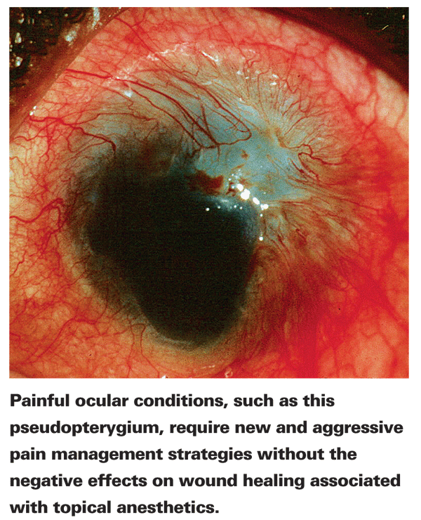
The central cornea is one of the most heavily innervated areas of the human body, with a nerve density about 600 times that of skin.13 These highly concentrated sensory peripheral nerve fibers are also known to contain opioid receptors, fueling speculation that topical treatment with an opioid agonist may provide effective pain relief and act as an alternative to widely used topical anesthetics.
Anesthesia, a term which comes from the Greek roots an- (without) and aisthesis (sensation), is inherently more dangerous than analgesia, derived from the roots an- (without) and algos- (pain). Unlike analgesics, anesthetics interfere with the body's natural defenses and wipe out sensations essential to the preservation and maintenance of tissue. In fact, prolonged treatment with all of the currently used ophthalmic anesthetics can cause damage to the corneal epithelium. These agents inhibit corneal re-epithelialization resulting in sloughing of the epithelium, alteration in lacrimation and mucous adherence. This process, in turn, leads to an instability of the tear film, increased corneal permeability and swelling, loss of corneal transparency, and alteration of the corneal cytoskeletal elements (actin and myosin), causing a disruption of the normal cell cycle.
In contrast to current topical anesthetics, opioid analgesics can alleviate pain following a corneal abrasion without a negative effect on corneal wound healing. Gholam Peyman, MD, and his colleagues were the first to document the effective use of topical opioids as analgesics in ophthalmic care in 1994.14 In their study, topical administration of morphine sulphate in patients with corneal abrasions provided analgesia with no negative effect on corneal wound healing.14 Conversely, repeated application of the non-opioid local anesthetic proxymetacaine 0.5% caused a significant delay in wound healing. Topical morphine sulphate didn't produce analgesia effects in patients with an intact cornea, suggesting that the analgesic effect may be due to activation of opioid receptors on inflamed tissues. In support of this concept, another research report using nerve fibers removed from the rat tibia showed that morphine decreases the activity of cutaneous nociceptors only under conditions of inflammation.15
A clinical study examining the effect of the topical mu-receptor agonist loperamide in cases of corneal abrasion, embedded foreign body and pterygium found the drug to be safe and marginally effective in reducing pain one hour after dosing.16 Loperamide, which is currently available over-the-counter as an anti-diarrheal drug, does not cross the blood-brain barrier and therefore lacks the central nervous system side effects and addictive potential of other opioids. In patients with corneal abrasions, analgesia was most pronounced in those who had experienced the trauma 48 or more hours prior to administration of the drug,16 a result that may have been due to a lag in receptor upregulation.
A more recently completed clinical trial assessing the analgesic effect of topical fentanyl (10 µg), a synthetic opioid agent, combined with dexpanthenol, a drug that improves epithelial wound healing, found no significant difference in pain relief after corneal surgery between those patients who received fentanyl and those who didn't receive the drug.8 The small concentrations of fentanyl may not be sufficient to penetrate pre-corneal barriers and reach the corneal epithelium, thus preventing the analgesic effects of opioid receptor activation. Furthermore, the topical fentanyl dose didn't produce systemically effective opioid blood levels which, by contrast, have been reached after the application of higher concentrations of topical morphine in one study.8
Corneal Wound Healing
The endogenous opioid known as opioid growth factor has been found to downregulate epithelial cell division and migration of cells in the closing of epithelial abrasions.17 OGF and the OGF receptor, a functionally unique opioid receptor that regulates cell division, have been identified in the cornea of the dog, cat and horse and are believed to inhibit the healing of damaged tissue in humans.17 OGFs are continually produced within cells of the cornea and conjunctiva where they bind to OGF receptors and inhibit DNA replication and cell proliferation.
Investigators recently found that inhibition of OGF activity with the use of gene therapy was shown to accelerate repair of the corneal epithelium, while increasing OGF through gene transfer delayed re-epithelialization.7 In a separate study, administration of the OGF antagonist naltrexone increased epithelial wound healing following corneal abrasions in the diabetic rat.4 These results suggest that OGF inhibition can help restore corneal cells in diabetic patients, who are at an increased risk of visual loss due to a delay in re-epithelialization following ocular trauma.7
IOP Reduction
Opioids, administered intravenously, periorbitally or topically, represent a novel pharmacologic option with initial research showing promising results in reducing IOP. Intravenous administration of heroin decreases IOP in rabbits, and morphine given intramuscularly has been shown to decrease eye pressure as well.18
Interestingly, another study found that patients addicted to either heroin or morphine had significantly lower IOPs than controls19 and that IOP increases following the administration of the opioid antagonist naloxone could be used to detect systemic opioid abuse.20
This research has lead to the speculation that topical, peripherally acting eyedrops may have a similar effect. In fact, topically administered opioids have been shown to reduce IOP in rabbit models. Mu-, delta- and kappa-opioid receptor agonists have all been shown to increase aqueous outflow.3,21 This increase in outflow can also be blocked by pretreatment with an opioid antagonist, although the precise mechanism by which these compounds affect IOP is not known.3
Unilateral topical application of dynorphin has been shown to cause a bilateral reduction in IOP beginning about one hour post-instillation, with a slightly larger effect in the treatment eye.3 A 1-µg dose produced a decrease of about 0.9 mmHg after an hour, while doses of 10, 33 and 100 µg resulted in maximum IOP reductions of about 3, 5 and 6 mmHg respectively. Differences between the treated and non-treated eye were most pronounced at lower dosages, suggesting both a systemic and localized effect.3
Researchers recently showed kappa-opioid agonist bremazocine (BRE) caused IOP reduction in monkeys. This IOP-lowering effect is postulated to be due to aqueous flow suppression via non-opioid receptor stimulation. High doses of BRE (100 µg) may cause mean arterial pressure to lower and subsequently cause substantial reductions in IOP. At lower doses (10 µg) a differential IOP-lowering effect is found in ipsilateral eyes, suggesting a more localized effect.22
Miosis
The most well-established effect of opioids on the eye is their influence on pupil diameter. Opioid administration typically causes miosis in humans, although rapid fluctuations in constriction and dilation are known to occur.3 This variation is thought to be caused by alterations in the transmission of multiple neurotransmitters. In the rabbit eye, it was shown that dynorphin, a relatively selective kappa-opioid receptor agonist, has dual effects on cholinergic and substance P-ergic transmission in the iris sphincter muscle, causing a dose-dependent constriction or dilation pupillary response.3 Dynorphin administered unilaterally at 100 µg caused miosis in the ipsilateral eye, while doses of 33 µg caused bilateral mydriasis.3
Opioid receptors are present on sympathetic nerves of the iris, and the pupillary effect has been largely attributed to changes within the oculomotor center, as unilateral topical application of an opioid agonist in monkeys was shown to cause bilateral pupil constriction.22 A pupil-constricting topical opioid may be useful for patients to quicken the return to normal visual function following a dilated ophthalmic exam.
Since opioids produce an analgesic effect without any unwanted effects on corneal
healing, it's worth pursuing the development of topical opioid analgesics that are more potent than non-steroidal anti-inflammatory drugs and can take the place of anesthetics in situations in which it's vital that wound healing isn't compromised. Also, the potential of these potent compounds not only as pain medications, but also as alternative IOP-lowering agents, makes it clear that viable, peripherally acting opioid formulations may not be too far away from clinical use. Opioid eyedrops have demonstrated clinical usefulness while avoiding the undesirable side effects associated with the systemic use of many other drugs. These future ophthalmic opioid formulations may effectively control pain, treat glaucoma and assist with corneal wound healing, effects that would demonstrate the amazing longevity and usefulness of these ancient drugs.
Dr. Abelson, an associate clinical professor of ophthalmology at
1. Aggrawal A. Narcotic Drugs.
2. Martin WR. Opioid antagonists. Pharmacol Rev 1967;19:4:463-521.
3. Russell KR,
4. Klocek MS, Sassani JW, McLaughlin PJ, Zagon IS. Topically applied naltrexone restores corneal reepithelialization in diabetic rats. J Ocul Pharmacol Ther 2007;23:2:89-102.
5. Stein C, Schafer M, Machelska H. Attacking pain at its source: New perspectives on opioids. Nat Med 2003;9:8:1003-8.
6. Monassier L, Bousquet P. Sigma receptors: From discovery to highlights of their implications in the cardiovascular system. Fundam Clin Pharmacol 2002;16:1:1-8.
7. Zagon IS, Jenkins JB, Sassani JW, et al. Naltrexone, an opioid antagonist, facilitates reepithelialization of the cornea in diabetic rat. Diabetes 2002;51:10:3055-62.
8. Zollner C, Mousa S, Klinger A, et al. Topical fentanyl in a randomized, double-blind study in patients with corneal damage. Clin J Pain 2008;24:8:690-6.
9. Husain S,
10. Gulur P, Weber D, Acquadro M. Anesthetics. In: Abelson M, ed. Principles and Practices of Ophthalmology, vol 1.
11. Celiker V, Basgul E, Sahin A, et al. Comparison of midazolam, propofol and fentanyl combinations for sedation and hemodynamic parameters in cataract extraction. Saudi Med J 2007;28:8:1198-203.
12. Nissman SA, Tractenberg RE, Babbar-Goel A, Pasternak JF. Oral gabapentin for the treatment of postoperative pain after photorefractive keratectomy. Am J Ophthalmol 2008;145:4:623-9.
13. Rozsa AJ, Beuerman RW. Density and organization of free nerve endings in the corneal epithelium of the rabbit. Pain 1982;14:2:105-20.
14. Peyman GA, Rahimy MH, Fernandes ML. Effects of morphine on corneal sensitivity and epithelial wound healing: Implications for topical ophthalmic analgesia. Br J Ophthalmol 1994;78:2:138-41.
15. Wenk HN, Brederson JD, Honda CN. Morphine directly inhibits nociceptors in inflamed skin. J Neurophysiol 2006;95:4:2083-97.
16. Nevius JM, Welch DL, Sandman E, Abelson M. Efficacy evaluation of ADL 2-1294 (loperamide hydrochloride ophthalmic solution 0.025%) vs. placebo for short term pain management. Invest Ophthalmol Vis Sci 2000(Suppl:4917).
17. Sassani JW, Zagon IS, McLaughlin PJ. Opioid growth factor modulation of corneal epithelium: Uppers and downers. Curr Eye Res 2003;26:5:249-62.
18. Green K. Ocular effects of diacetyl morphine and lysergic acid diethylamide in rabbit. Invest Ophthalmol 1975;14:4:325-9.
19. Drago F, Panissidi G, Bellomio F, et al. Effects of opiates and opioids on intraocular pressure of rabbits and humans. Clin Exp Pharmacol Physiol 1985;12:2:107-13.
20. Drago F, Aguglia E, Dal Bello A, et al. Ocular instillation of naloxone increases intraocular pressure in morphine-addicted patients: A possible test for detecting misuse of morphine. Experientia 1985;41:2:266-7.
21.
22. Rasmussen CA, Gabelt BT, Kaufman PL. Aqueous humor dynamics in monkeys in response to the kappa opioid agonist bremazocine. Trans Am Ophthalmol Soc 2007;105:225-38; discussion 38-9.


