New research, which sought to gain a better understanding of the prevalence of Acanthamoeba keratitis, found that it has a relatively low incidence in the general population but it remains strongly linked with contact lens wear.
While Acanthamoeba keratitis is a significant cause of infectious keratitis, a comprehensive assessment of the incidence of this condition remains lacking. To address this gap, an international group of researchers (representing institutions in the U.S., U.K., China and Italy) initiated a systematic review and meta-analysis to examine the incidence of AK using the currently available data from peer-reviewed literature.
In this analysis, researchers calculated the incidence of AK as the number of cases per health-care center per year (annualized-center-incidence). They also calculated the following meta-analytical ratios: (a) the ratio of AK eyes to the count of non-viral microbial keratitis (MK) eyes, and (b) the ratio of AK eyes to the overall population (i.e., the total number of subjects of a nation or region, as indicated by the authors in each study).
Overall, the study included 105 articles published between 1987 and 2022. Investigators identified a total of 91,951 eyes, with 5,660 affected by AK and 86,291 by non-viral microbial keratitis. Data showed that the median annualized-center-incidence was 19.9 new Acanthamoeba keratitis eyes per health-care center per year, with no statistically significant differences observed among continents. Risk factors did, however. “Acanthamoeba keratitis, in regions with high per capita incomes, primarily affects contact lens wearers,” the authors wrote, “whereas elsewhere ocular trauma, often in agricultural workers, is the major association.”
Additionally, the study authors found that the ratio of AK eyes to the total number of MK eyes was 1.52 percent. Comparatively, the ratio of AK in relation to the entire population was estimated at 0.0002%, or 2.34 eyes per 1,000,000 subjects, according to the study authors.
“Our analysis demonstrates AK as a relatively low-incident condition among the general population, with no major differences in terms of incidence among different continents,” the study authors wrote.
“These studies will provide valuable insights for improving our understanding of Acanthamoeba keratitis epidemiology and implementing effective strategies for prevention, diagnosis and management,” the research team concluded.
1. Aiello F, Afflitto GG, Ceccarelli F, et al. Perspectives on the incidence of Acanthamoeba Keratitis: A Systematic Review and Meta-Analysis. Ophthalmology. August 8, 2024 [Epub ahead of print].
Higher-order Aberrations in Fuchs’ Patients Studied
A new analysis showed significant changes in high-order aberrations among Fuchs’ endothelial corneal dystrophy patients with subclinical corneal edema. This data, recently published in the journal Cornea, highlight how tomographic analysis can support visual impairment assessment, disease progression and decision-making for early endothelial keratoplasty in these individuals.
This retrospective, single-center case series, which analyzed whether tomographic changes in Fuchs’ dystrophy impact the higher-order aberrations in the early period of the disease, included a total of 78 eyes of 47 patients with Fuchs’ endothelial dystrophy with slit-lamp biomicroscopically visible guttae, but no visible corneal edema. These patients were assigned to group 0 (no subclinical corneal edema) or 1 (subclinical corneal edema).
Group 1 was composed of 50 eyes of 33 patients with a mean age of 70.9 years and group 0 included 28 eyes from 18 patients with a mean age of 63.9 years. In group 0 and 1, 13 eyes and 34 eyes were pseudophakic, respectively.
Sixty-six healthy eyes of 38 patients were also included in the analysis as the control group. Of these, 58 eyes were phakic and eight were pseudophakic.
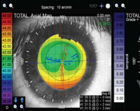
Study authors calculated mean values and standard deviations for the root mean square (RMS), coma, trefoil and spherical aberrations of the cornea, the anterior surface and the posterior surface.
Data demonstrated statistically significant differences in the RMS higher-order aberrations, coma and spherical aberrations of the posterior surface in group 1 eyes when compared to those in group 0. No statistically significant differences in high-order aberrations were observed between the control group and eyes without subclinical corneal edema (Group 0).
These findings showed statistically significant increases in high-order aberrations, particularly of the posterior corneal surface, in patients affected by Fuchs’ endothelial dystrophy with subclinical corneal edema, according to the study authors, who noted, “Our study adds to the current literature by providing evidence of significant changes in high-order aberrations in patients with Fuchs’ endothelial corneal dystrophy with subclinical corneal edema compared with those without.
“Early tomographic analysis can, therefore, aid in detecting subclinical corneal edema with varying magnitude of high-order aberrations compared with patients with Fuchs’ endothelial dystrophy without subclinical edema or healthy eyes,” the authors added, “which, in turn, can help characterize the patients’ visual impairment and decision-making for early endothelial keratoplasty.”
1. Blöck L, Son HS, Köppe MK, et al. Corneal high-order aberrations in Fuchs’ endothelial corneal dystrophy and subclinical corneal edema. Cornea. July 30, 2024 [Epub ahead of print].


