Current treatment for neovascular age-related macular degeneration includes intravitreal injections of bevacizumab (Avastin) or ranibizumab (Lucentis), every four weeks. Lucentis is approved by the Food and Drug Administration for the treatment of AMD; Avastin is currently used off-label.
Epimacular brachytherapy (Neo- Vista) is a novel treatment for neovascular AMD that may reduce the number of anti-VEGF intravitreal injections required and may further improve visual acuity. This treatment consists of applying beta radiation in a very selective, focused way so that patients experience the benefits of radiation, with minimal damage to collateral tissue.
Pravin U. Dugel, MD, is an investigator in MERITAGE-I, an ongoing multicenter international clinical study evaluating the safety and efficacy of epimacular brachytherapy. "With this new treatment, we want to do two things: one is to decrease the frequency of treatment, and the other is to improve the efficacy of treatment by targeting multiple pathologic pathways involved in the disease. Years ago, it was shown that external beam radiation had an effect that was beneficial; however, due to the large area of exposure to the radiation applied, there was substantial damage to the surrounding tissues, which led to a number of side effects," says Dr. Dugel, who is a managing partner at Retina Consultants of Arizona in Phoenix.
The beta radiation treatment is administered in the epimacular region targeting the neovascular membrane, after performing a core vitrectomy. "Once the vitreous is removed, the small tip of the radiation device is inserted in the eye and placed strategically right over the neovascular membrane," says Dr. Dugel. "It is held there for about four minutes, and then removed. Nothing is left in the eye."
Using a strontium 90 beta isotope, the focused energy is delivered to a target area up to 5.4 mm in diameter. Importantly, patients' systemic exposure to radiation is minimal. In fact, the dose to the entire body during this procedure is less than that from a standard chest X-ray.
Although it is a surgical procedure with all the inherent risks, the risk of any radiation toxicity is minimal. "Performing a vitrectomy prior to administering the therapy may have beneficial effects," says Dr. Dugel. "Just doing the vitrectomy increases the amount of oxygen in the back of the eye, which enhances the effect of beta radiation on the neovascular tissue. The process also involves giving an anti-VEGF injection at the time of the procedure, if required, to inhibit any acute change in VEGF expression."
The preliminary study results were recently presented at the AAO's annual meeting as part of the late-breaking news section. This study included patients who had as many as 23 injections and more than two years of anti- VEGF therapy before receiving epimacular brachytherapy. All patients were required to have received a minimum of eight injections during the past 12 months or six injections in the past six months preceding enrollment. Sixteen patients were included in the preliminary study results, and these results suggest that a single epimacular brachytherapy procedure can further improve visual acuity in this patient population while decreasing the number of anti-VEGF injections required. Interestingly, 63 percent of patients experienced an improvement in their visual acuity, and 50 percent gained five or more letters of visual acuity at six months.
This ongoing, multicenter study has completed the targeted enrollment of 50 patients at two centers in the United States, one in the UK, and two in Israel. A limited number of adverse events have occurred to date. These include subconjunctival hemorrhage, vitreous hemorrhage and cataract formation. All are related to the vitrectomy procedure, not the epimacular brachytherapy. To date, no instances of radiation toxicity have been reported.
"Unlike Avastin and Lucentis, epimacular brachytherapy does more than just stop angiogenesis," says Dr. Dugel. "It offers a broad spectrum of activity and may therefore inhibit the multiple disease processes involved in the pathology of neovascular AMD. There is existing evidence in the oncology world to show that combination therapy with radiation and anti-VEGF agents has optimal clinical outcomes."
NeoVista has recently completed enrollment for a pivotal trial called CABERNET (CNV Secondary to AMD Treated with Beta Radiation Epiretinal Therapy). This multi-center, randomized, controlled clinical study has enrolled more than 450 subjects at 45 sites worldwide and is evaluating the safety and efficacy of epimacular brachytherapy performed concomitantly with Lucentis compared with Lucentis therapy alone.
The commercial product Vidion ANV therapy system was CE marked in mid-2009, and sales have already begun in Europe. Commercial activity surrounding epimacular brachytherapy will begin in the United States in 2011, pending FDA approval.
NDA Submitted for Once-Daily Post-Cataract Drug
Ista Pharmaceuticals has submitted a supplemental New Drug Application to the Food and Drug Administration for bromfenac ophthalmic solution dosed once-daily as a treatment for ocular inflammation and pain following cataract surgery. Ista currently markets Xibrom (bromfenac ophthalmic solution) 0.09%, which is an eyedrop labeled for use twice-daily, beginning 24 hours after cataract surgery. If approved, Ista plans to market the once-daily product under the brand name XiDay.
Ista filed the XiDay NDA electronically and pending FDA validation of the electronic file and timely acceptance of the filing by the FDA, the company expects a standard review of six months from date of receipt.
Florida Center Combines Eye Banking and Research
With the October 2009 opening of the Lions Eye Institute for Transplant & Research, the first of its kind to be physically connected to an eye bank, researchers in ocular disease now have increased access to fresh corneal tissue samples almost immediately after they are removed from donors, a difficult task in the past.
The Tampa, Fla.-based LEITR began developing this new design for an ocular research center in 2007 after deciding to shift efforts toward not just procurement and transplantation but disease research. "The bigger picture isn't transplantation, it is eye disease," says Jason Woody, research CEO for LEITR. "Eye diseases such as glaucoma, macular degeneration and diabetic retinopathy affect millions of people, and if we can help to move toward cures and treatments along those areas, that would obviously make a bigger splash in the pond." Mr. Woody explains that the goal of LEITR—which was founded in 1973 by a group of Tampa Lions dedicated to fighting blindness—was to create a new method of ocular research that would "fast-track the pharmaceutical companies' researchers into working at a quicker pace. The idea is that if we could bring the availability of viable research tissue to researchers it would move things forward and force the pharmaceutical companies that may be working toward a similar goal to take a look at that and compete by moving their research forward as well."
According to Mr. Woody, the availability of viable research tissue often stands as a roadblock for many researchers. By providing a readily available source of tissue that has been removed from donors as recently as 12 to 24 hours prior to use, researchers can evaluate disease and treatments in tissue much more similar to that found in functional eyes, as opposed to the tissue most researchers may be used to, which has been altered due to transport and the passage of time.
The initial platform of the facility will be AMD and glaucoma research. The Ocular Research Center measures 12,000 square-feet that is occupied not only with lab space, but also dormitories for out-of-state researchers.
Since its inception 30 years ago, the organization has provided more than 50,000 eyes for research. With the Ocular Research Center, LEITR plans to use its facilities to offer new options to investigators who often had to settle for animal models or poor tissue samples that could prevent them from fully understanding how disease affects the eye. Along with being able to provide higherquality tissue samples, LEITR's Ocular Research Center's adjacent eye bank can also provide researchers with a much more thorough background and medical history as well, providing a fuller understanding of diseases and their effects.
"We really don't have any good animal models that we can use to recreate conditions like AMD," says Henry Edelhauser, PhD, of Emory Eye Center in Atlanta, a board member for LEITR. With the abundance of readily available tissue at their disposal, Dr. Edelhauser feels that researchers will be able to study these conditions in a new light. "With the eye bank attached, there is now this great capability to acquire fresh tissue for various types of analysis so we can understand what the different stages of these diseases in the human eye actually are. Once we can understand the different stages on the tissue level and even the molecular level, then we can really start to think about how to use therapeutic agents for better treatment." 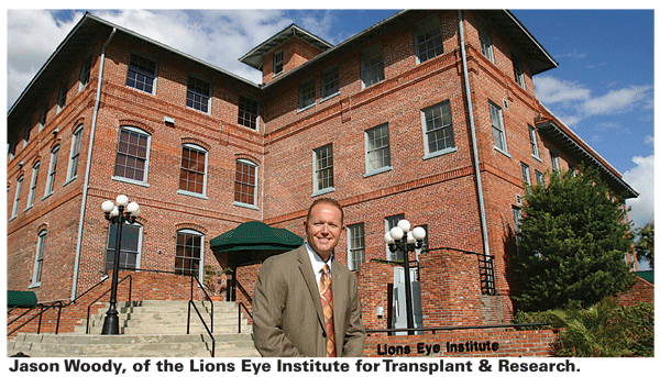
LEITR is able to take full advantage of Florida's elderly populace—many of whom suffer from some of the conditions being studied—as well as the state's high mortality rate, giving the organization a wealth of tissue and the ability to offer researchers approximately three to five pairs of fresh eyes per day. With so many donor eyes at its disposal, the new facility will also provide a means to using the whole donated eye, and not just the corneal tissue, for research advancements.
"We currently don't have a way to transplant the retina, but we can still use that tissue for various studies," explains Los Angles-based surgeon, Mark Humayun, MD, PhD, who also serves as a board member for LEITR. "If you can find a way to preserve the whole eye, then there are other things you can use that for other than just simple procurement."
"Many investigators around the United States do not have access to human research tissue, so the new facility can provide research tissue to investigators across the country," says Dr. Edelhauser. "The key to all this is to be able to provide the tissue relatively fresh, so that biochemical, physiological and molecular analysis can be accurately done."
For information, visit lionseyeinstitute.org.


