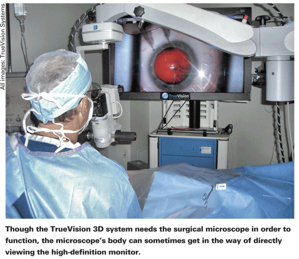
The surgical microscope is an indispensible part of ocular surgery, and will be for the foreseeable future. However, for many surgeons, having to hold the same physical position—leaning over the oculars for an extended period of time can become onerous—and sometimes even unhealthy for the neck and back. TrueVision Systems is trying to give surgeons a new way to operate by way of a high-definition 3D monitor that gets its image from a camera located on the surgical microscope. Here's a look at the system, its pros and cons, and the features it might offer in the future.
A Period of Adjustment
The TrueVision system consists of a high-definition camera that's attached to the surgical microscope, which highlights the fact that the technology isn't at the point where it will replace the surgical microscope altogether. The camera is connected to a powerful computer processor by a cord that runs up and over the microscope. The data comes out of the processor and is fed to a large, flat-panel HD monitor that displays the surgery at a resolution of 1080p.
Eugene
Also, in the original system, there was a time lag between when I'd move an instrument and when I'd see it move on the screen. It was only a few milliseconds, but it was noticeable. That was eliminated when the company increased the processor speed."
 Dr. Packer says it's not something you can just sit down and begin using immediately, though. "I encourage surgeons who get the system to do a dry run," he says. "Maybe have a nurse lie down so you can figure out the positioning, which is key. Once you've got the physical setup in place, you then have to use the software to control the system.
You've got options for adjusting such things as color, contrast, brightness and aspect ratio. Once you get the image the way you want it, you can lock in your settings. Sitting down with your first case isn't the best time to do all of this."
In terms of color settings, for instance, Dr. Packer says you can set up the system to mimic what you see through the microscope based on a particular kind of illumination, such as xenon or halogen. "Or you can make it different," he says. "I like to enhance the red reflex a little bit to give it a deep, sunset-red look. It really highlights the red reflex.
When I use fluorescein dye at the end of the case to make sure the wound isn't leaking, the dye shows as blood red on the screen. It's surprising when you see it for the first time if you're used to the yellow-orange color of fluorescein. But, for every setting you choose, something else might have to be compromised. With my setting, I lose a little illumination on the conjunctival surface, but that's not critical to me."
In terms of getting used to the system, David Friess, OD, TrueVision's director of clinical and regulatory affairs, says first-time TrueVision surgeons will find themselves using the microscope's oculars at times. "For the first case, the surgeon will use the oculars and then use the screen off and on for certain portions of the procedure," he says. "Then, he'll progressively move toward looking at the screen more often during a handful of initial cases. It's kind of comforting being able to move back and forth between the oculars and the screen as needed. Often, surgeons will be performing most of their cases from the screen after six or eight surgeries."
The Screen vs. the Scope

At present, the main benefit of the TrueVision system is ergonomic. Using the screen frees the surgeon from bending over the microscope case after case. "It's more comfortable looking at a screen," says Dr. Packer. "It's less stressful physically. You don't have to keep your eyes in a certain place, and instead can just lean back rather than having to keep leaning over. Your head is in a normal position."
However, looking at the screen isn't the same as viewing the surgery through a microscope's oculars. "One thing you'll notice, because it's a digital image, is that, even though it's HD, it doesn't have the infinite resolution that you'd get by looking through an optically optimal set of lenses and using your eyes. It's kind of like the difference between reality and the high-definition image on your TV set. It's perfectly adequate for cataract surgery, though. You can see strands of cortex and individual lens epithelial cells on the anterior capsule. You can still see more than you need to see, but it's just you're not seeing as much as you may be used to seeing. So that's kind of a trade-off.
For instance, I think corneal endothelial cells would be difficult to see with the system, even under the highest magnification, whereas I can just make out the edges of the endothelial cells when looking through my Zeiss Lumera microscope under high magnification. However, there's nothing I feel that I can't do in cataract surgery with it.
I've done small-pupil IFIS cases, put in capsular tension rings and sutured in lenses. I primarily use it for cataract surgery, but it's perfectly fine for glaucoma surgery, too. I tried using it for plastics, such as lid procedures, but the microscope field of view is too small. It can't zoom out enough to include enough of the face on the monitor, so I still use loupes for oculoplastics cases."
 The system also needs a certain level of illumination to be viewable. "If the room lights are too bright, that decreases your ability to see the screen," says Dr. Packer. "So you have to turn them down. However, your scrub nurse still needs some light to see the instruments, so we use a ceiling-mounted operating light to illuminate the scrub table."
Dr. Packer says the system's 3D view does give a better depth of field than just looking through the microscope, however. "You have tremendous depth of field," he says. "I don't focus up and down much using the system. I think at the very beginning of the case I try to maximize my focus on the cornea and the anterior capsule, and may focus down a bit more during phaco, but I often just leave it alone."
The system is also expensive, which will limit the number of surgeons who will jump into using it. "Because of the cost, we have one, not two, for our two operating rooms," says Dr. Packer. "We have a TrueVision system in one OR and a surgical microscope in the other." The system costs $75,000, but, as was mentioned earlier, it also requires a surgical microscope to use.
For $24,000 extra, surgeons can purchase a digital recording and editing software suite for PC or Apple computers called TrueEditor that gives surgeons the rudiments necessary to edit 3D surgical videos for presentation or educational purposes. "We saw a big need for a really simple editor," says Burton Tripathi, PhD, TrueVision's vice president of product development, "something that lets you set your cut points and establish your final edited film in either 3D or 2D. TrueEditor has scene transitions like fades and wipes, and surgeons can put annotations on the screen."
Future Directions
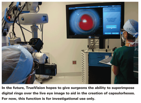
The next step for the TrueVision system is the use of digital guidance overlays on the 3D image to aid surgeons in making capsulorhexes and limbal relaxing incisions and implanting toric intraocular lenses. The company hopes the addition of such features will help the system actually make an impact on outcomes, in addition to improving ergonomics.
"For the guidance systems, we use the 3D camera on a slit lamp to get a 3D still image, and then import that still image into the OR system," explains Dr. Friess. "In the OR, the surgeon brings up the still image on the monitor and registers it with the live 3D view of the eye using anatomic landmarks. When the two images are registered together, the surgeon can place a digital overlay on the live image to help with steps of cataract surgery. For instance, to place LRI incisions, the surgeon will be able to look at the screen in the OR and follow the markings overlaid on the eye." The digital guidance overlays for capsulorhexes and LRIs have been submitted to the Food and Drug Administration for 510(k) approval, and the study into overlays for toric intraocular lens placement is just beginning.
Also, the ultimate goal of the system is to go completely digital and do away with the need for a microscope. "One of the things that's still a little cumbersome is the fact that you have to use a microscope," opines Dr. Packer. "The scope is kind of in the way. I know the big wish is to just have a camera with a lens system that can focus on the eye that's all digital."
The company isn't sharing a lot of details about how close such a system is to reality, however. "The potential is there," says Dr. Friess.
In the meantime, surgeons who are growing weary of using the surgical microscope for all their cases, or who would like a way to produce new, 3D teaching videos might think about giving the system a try. "The ergonomic benefit is key and the educational benefits of being able to present 3D videos at meetings offer a new learning opportunity for colleagues," says Dr. Packer. "I think those two things are the biggest selling points of the system."


