As simple as it seems, the question in the headline above is the standard that often constitutes successful cataract surgery in veterinary ophthalmology. Don't apply to veterinary school just yet, though! There are differences and difficulties we veterinarians encounter with cataract surgery that would send you running back to your HMO woes. In this article, I'll offer a veterinary ophthalmologist's perspective on some challenges and rewards that we share with you, and those that are unique to the canine branch of eye care.
Canine Cataract
The majority of cataracts that occur in younger dogs are inherited cataracts, and there is a long list of affected breeds. This list includes the Poodle, Labrador and Golden Retriever, Siberian Husky, American Cocker Spaniel, Bichon Frise and Miniature Schnauzer. Other causes of canine cataract formation include diabetes mellitus, uveitis, trauma, toxins, nutritional factors or irradiation. Nuclear sclerosis is usually clinically evident by 6 years of age, though it is not a common cause of lens surgery. Cataract surgery is usually, but not exclusively, performed when the dog shows demonstrable visual impairment, such as not able to find toys or bumping into furniture.
The canine lens is larger than the human lens; measuring approximately 7 mm from anterior to posterior pole with a volume of 0.5 ml. Equatorial diameter is 10 mm. The average aphakic dog requires a 41 D intraocular lens implant for correction. The canine lens capsule is 50-70 µm thick anteriorly and 2-4 µm thick posteriorly.
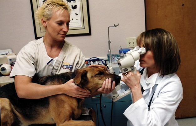 |
| Dr. Christi Warren examines Teppo, a cataract patient, with an assist from technician Kristina Wirths. (All images Robert Walker Photography.) |
Our canine patients require rigorous medication schedules of topical and systemic anti-inflammatory medications, prior to surgery to control lens-induced uveitis and afterwards to control postoperative inflammation. Medications are often continued long-term to control postoperative uveitis, which is more severe and longer lasting than what occurs in the average human cataract patient.
Surgical Considerations
To undergo cataract surgery, our patients are fully anesthetized. The patient is intubated and maintained on Isoflurane. I use atracurium for paralyzation, which allows for easy globe positioning and manipulation. The patients are ventilated and CO2, O2, EKG, BP, and other vital parameters are monitored. The positioning of the patient can be tricky, as we have to deal with a large range of head and nose sizes and conformation. The more easily positioned dogs tend to be the brachycephalic breeds such as the Pugs and Lhasa Apsos because of their short nose length and prominent globes. Compare this with a 90-pound Golden Retriever with a much larger nose and deeply recessed globes. In addition, we must contend with the dog's nictitating membrane, or third eyelid, located nasally in the palpebral space, which seemingly gets in the way of everything during surgery.
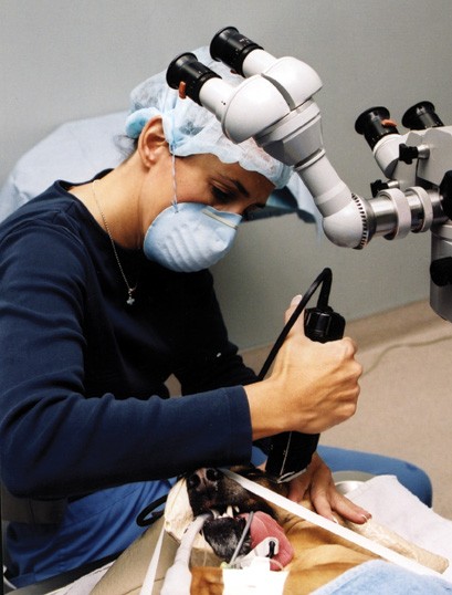 |
| Sue Frey, another technician at Eye Care for Animals–Tucson preps the patient for surgery. Postop uveitis is a major concern with canine patients. |
Most veterinary surgeons perform perilimbal clear corneal incisions. The canine sclera is highly vascular and the chance of hemorrhage and iris prolapse is increased with scleral incisions. Therefore scleral tunneling is not a technique utilized in canine cataract surgery.
The thicker anterior lens capsule does not lend itself well to capsulorhexis with a cystotome. Instead, a circular capsulorhexis is performed with an initial stab incision through the capsule, and the flap is grasped with a forcep (i.e., Utrata forcep) accomplishing a circular tear. Hydrodissection is also more difficult because canine cortical material is very thick and tenacious. I find that the broad-fanned stream of the Chang hydrodissection cannula works well in our canine patients. Great care must be taken to not touch the iris at any time, making good pupil dilation critical. The slightest touch of the iris can result in posterior synechiae or hemorrhage.
Issues in Technique
Veterinary ophthalmologists utilize phacoemulsification for lens removal. Our machines must be powerful to deal with the hard canine lenses, and I use the AMO Sovereign. Most surgeons utilize a one-handed technique, though I prefer a two-handed horizontal phaco chop. Katena has manufactured a canine chopper which is a 4-mm long microfinger (Fishkind-Warren chopper) modified from the Chang chopper. The longer chopper allows for a more complete chop in these 7-mm thick canine lenses. Since many of our patients are presented for surgery when the cataract is in an opaque mature stage, I often have to chop the entire lens, as there is little distinction between nucleus and epinucleus. This means reaching farther under the capsule to the equator with the chopper to get around the lens. The leftover cortex is thick and sticky, and capsular cleanup is difficult. We utilize the largest port available on our I/A tip.
| A Great Outcome for a Horned Owl | |
| Veterinary ophthalmology won some rare but widespread attention with an Associated Press wire story that many major consumer media outlets picked up back in February. After wildlife rehabilitators in Wisconsin were alerted that a great horned owl had been sitting on a fence in the same field for several days, they captured the bird and discovered that it had cataracts. The bird was brought to the University of Wisconsin-Madison School of Veterinary Medicine. There, Chris Murphy, DVM, PhD, a veterinary ophthalmologist, and his colleagues performed cataract surgery and implanted intraocular lenses, telling the AP that they believe it was a first for such a bird. Dr. Smith says that great horned owls do develop juvenile cataracts, though he was uncertain as to the etiology in th 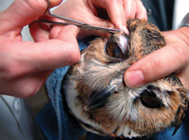 is case. Experienced in raptor ophthalmology, Dr. Smith says that raptors in the wild have a very high frequency of eye disease and lesions, though the great majority are due to trauma resulting from some type of contact with humans, such as hunting or entrapment. is case. Experienced in raptor ophthalmology, Dr. Smith says that raptors in the wild have a very high frequency of eye disease and lesions, though the great majority are due to trauma resulting from some type of contact with humans, such as hunting or entrapment.The owl eye, as do those of many birds, contains striated muscles that preclude the use of a muscarinic. Instead a curare form of drug was injected intracamerally to dilate the pupil for the procedure. Scleral ossicles, or bones, also distinguish the ocular anatomy and dictate a clear corneal incision. In addition, the animal anatomy prevents the use of the formulas commonly used to calculate IOL power in humans, though Dr. Smith says that their use of more traditional methods, variations on the Binkhorst equation, was accurate enough that the bird was within a quarter diopter of emmetropia postoperatively. One area where owls are similar to humans, he adds, is their relatively low level of postop inflammation following cataract surgery. Other species, such as dogs and especially horses, are far more prone to complications from inflammation. Dr. Smith reports that the bird's recovery has been "perfect," and she was scheduled for release at the end of April. —Chris Glenn, Editor in Chief | |
IOL implantation is performed after cataract extraction. Lenses are manufactured specifically for the dog, and PMMA and acrylic lenses are available. I utilize the canine foldable in-the-bag lens of 41 D and 12-14 mm in diameter made of a hydrophilic acrylic inserted through a 3-mm incision.
Postop
Postop complications include glaucoma, synechiae, retinal detachment, chronic uveitis and endophthalmitis. Complications can occur months to years after surgery. The success rate of canine cataract surgery is approximately 95 percent short-term (four to six weeks) but decreases to 70-85 percent long-term (six months and longer).
My veterinary ophthalmic practice is similar to a pediatric practice, and I care about every single patient as one would a child. So I am especially pleased when a cataract surgery patient, so visually impaired prior to surgery that he had to be carried into the exam room, returns for a postop recheck, and bounds through the exam room door, running to me to get doggie treats from my hand and cover my face with kisses. Oh, yes, he can now find his food bowl!
Dr. Warren is board-certified in veterinary ophthalmology and a diplomate of the American College of Veterinary Ophthalmologists. She received her veterinary degree from Texas A&M University and completed a three-year residency in veterinary ophthalmology at the University of Florida. She is a partner in Eye Care for Animals–Tucson. Contact her at: 141 E Ft Lowell Rd. Tucson, AZ 85705. Phone: (520) 888-4498 and e-mail: azdryeye@qwest.net.


