When it comes to using mitomycin-C as an adjunct to PRK for haze prophylaxis, I was under the impression that the surgeons who were going to use it already were doing so, and the ones who weren't had made up their minds not to. However, after speaking to surgeons at recent meetings, both from the podium and in the hallway, I'm struck by how many surgeons are interested in the drug but just can't decide one way or the other.
There are many physicians who have questions about how to begin using it as well as the best way to use it for various patient scenarios. Whichever camp you belong to, the current users or the merely curious, here are my impressions on the safety of mitomycin-C as well as suggestions on the best ways to use it in different situations.
Is It Safe?
Here's a look at some of the research on the subject of mitomycin-C toxicity, as well as my experience.
A prospective, randomized study from Mexico that used the fellow eye as a control and used 0.02% mitomycin-C for 30 seconds found that the endothelial cell loss was statistically significant in the mitomycin-C group at one and three months postop.1
Another study divided 81 patients with low to moderate myopia (162 eyes) into three groups: bilateral mitomycin-C application; unilateral and untreated. Overall, 76 eyes were treated with the drug. Six months postop, the researchers say they found a decrease in endothelial cell density (-14.8 percent) that was statistically significant compared to control eyes (-5.1 percent). The investigators report that longer drug contact time and male sex were associated with greater cell loss.2
However, there have also been a number of studies that have not found a connection between the drug and cell loss. Last year, in a prospective study from
Our group undertook a study of PRK with prophylactic mitomycin-C and found no significant effect on the endothelium. To standardize things as much as possible, and to control for any errors in counting the cells, we had the patients' specular microscopy images sent to an independent reading center for counting. The mean endothelial cell density before surgery was 2,882 cells/mm2, and after surgery it was 2,867 (p>0.05).4 In addition, the other major parameters of endothelial function (Coefficient of Variation and percentage of hexagonal cells) showed no change between the two groups.
Since endothelial cell studies can be variable based on how the cells are counted, I think it's important to have an independent reading center do the counting.
In addition to studies that haven't found mitomycin-C to be toxic, I think it's significant that we've been able to use the drug safely for about 13 years. I believe if there were going to be problems with our mitomycin-C regimen, we would have seen a consistent and progressive incidence of complications.
Putting It to Use
Though I feel mitomycin-C is safe, I still respect certain rules and guidelines for its use.
The question I hear the most deals with the indications for mitomycin-C in refractive-surgery patients. First, it's important to note that I've never recommended using it on every PRK patient, though some do. My main criterion for using it is that the patient should be at higher risk for haze—specifically, a patient in whom you're doing an ablation of 75 µm or deeper. Our typical usage protocol is 12 seconds of application of 0.02% mitomycin-C, then rinsing of the area with at least 30 cc of BSS.
Another very good question that has arisen lately is about the use of mitomycin-C in patients with high astigmatism who may not be undergoing a deep ablation. I've seen patients with high amounts of astigmatism whose ablation depth will be shallower than 75 µm but, because I'm concentrating the laser energy in a specific bow-tie distribution to treat the astigmatism, they have a higher risk for haze over that area. The energy will be particularly high in the periphery. Anecdotally, I've found that these patients can get scarring, and it's always in the area where the laser was concentrated the most. Though they won't experience the same vision issues that they would if the haze were central, they may get regression. That regression will lead to more treatment being required, which runs the risk for more haze. In cases of high astigmatism, then, which I would define as 2.50 D or greater, I generally recommend the use of mitomycin-C.
Another question some surgeons wonder about it is the efficacy of using an even lower dose of mitomycin-C: 0.002% vs. 0.02%. The Cleveland Clinic's Ron Krueger, MD, has studied this in detail, and found significantly less haze with the standard-dose group in higher myopia (>-6 D) and higher ablation depth (>75 µm) at all postop time points compared to the lower dose. Overall, it appears the standard dose is probably preferable.5
The other issue that surgeons have to deal with is how to approach treating haze after refractive surgery.
Anecdotally, it seems that whenever keratocytes are active and producing scar tissue, then the 12-second prophylactic dose isn't as effective. So, what I recommend for this type of patient is to use the drug for a two-minute application time.
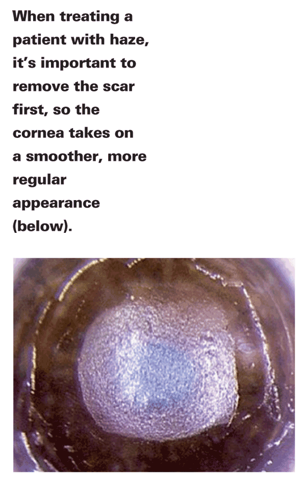
However, before you use the mitomycin-C, you have to remove the scar. The way I recommend removing it is either with the excimer in PTK mode or with a diamond-dusted pterygium burr, which you may be able to control a little better than a PTK. If I'm using the laser, I'll set it for the maximum spot size, 6.5 mm, and also program it for the maximum number of laser pulses allowed by the laser's key card (but I won't necessarily use all the pulses). A combination of the two may be ideal, however.
To eliminate the scar with either method, you have to avoid removing too much tissue and inducing a hyperopic refractive shift. To avoid this problem, I'll remove some of the scar and then take the patient to the slit lamp to examine the cornea. Typically, it takes two or three trips to the slit lamp to ensure the treatment's complete. You'll be able to see your progress very clearly at the slit lamp, but even under the laser microscope, you may be able to get a sense of the surface regularity; it will look a lot smoother when you're finished.
You can never get rid of the entire scar, but you should attempt to get rid of as much of it as you can. Once you feel you've removed most of the scar, apply the 0.02% mitomycin-C for two minutes. Most patients heal pretty quickly, and we haven't seen any cases of delayed re-epithelialization. Once patients have healed, their vision will usually be improved by a couple of lines, and most are fairly happy with the result.
What other options are there for haze treatment and prophylaxis? According to the literature, topical thiotepa is a possibility. In one study of the drug, researchers used it to treat five post-PRK patients with recurrent haze. Before treatment, the best-corrected vision ranged from 20/40 to 20/200. Afterward, three patients saw 20/20(-2) or better uncorrected and 20/20(-1) or better with correction. The other two eyes saw 20/25 best-corrected.6 I have no personal experience using the drug, however.
Dr. Majmudar is an associate professor of ophthalmology at
1. Morales AJ, Zadok D, Mora-Retana R, et al. Intraoperativemitomycin and corneal endothelium after photorefractive keratectomy. Am J Ophthalmol 2006;142:3:400-4.
2. Nassiri N, Farahangiz S, Rahnavardi M, et al. Corneal endothelial cell injury induced by MMC in PRK: Nonrandomized controlled trial. J Cataract Refract Surg 2008;34:6:902-8.
3. Leccisotti A. Mitomycin-C in hyperopic PRK. J Cataract Refract Surg 2009;35:4:682-7.
4. Goldsberry DH, Epstein RJ, Majmudar PA, et al. Effect of mitomycin C on the corneal endothelium when used for corneal subepithelial haze prophylaxis following photorefractive keratectomy. J Refract Surg 2007;23:7:724-7.
5. Thornton I, Xu M, Krueger RR. Comparison of standard (0.02%) and low dose (0.002%) MMC in the prevention of corneal haze following surface ablation for myopia. J Cat Refract Surg 2008;24:1:S68-76.
6. Anderson Penno E, Braun DA, Kamal A, et al. Topical thiotepa treatment for recurrent corneal haze after photorefractive keratectomy. J Cataract Refract Surg 2003 Aug;29:8:1537-42.