As the relentless shift toward digital technology in medicine continues, ophthalmology has followed suit—slowly. Now Clarity Medical Systems in
Keith Mullowney, president and CEO of Clarity, describes the P2 as the first fully digital integrated diagnostic and therapeutic station, combining multiple functions and systems with 3-D imaging and information storage. "Ophthalmic instruments and diagnostic tools are usually single-function and often analog," he notes. "In most cases, an eye exam is documented with a drawing of the eye and notes are manually transcribed into the patient's chart. This creates efficiency issues because the physician spends a lot of time acquiring and writing or transcribing the information. Also, the quality and accuracy of the transcription can be imperfect.
"Imaging and photography are helpful, but they're very skill intensive," he continues. "Stereo photographs typically require an experienced photographer; you'll probably have to move the patient to a station for the imaging to be done; and you may also have to dilate the patient."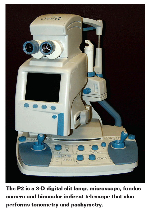
Mr. Mullowney notes that the P2 addresses these issues. "The P2 is totally digital," he says. "It's a 3-D digital slit lamp, microscope, binocular indirect telescope and fundus camera that also performs tonometry and pachymetry. It not only serves as a real-time display and measuring device, it can also record all of the information digitally—including 3-D images of the eye, exactly as the you see them. Yet it's only about half the size and weight of a traditional fundus camera, so it can fit on a tabletop or in the exam lane."
The Advantages of 3-D
"Most imaging systems in use today are still 2-D," Mr. Mullowney says. "However, the two traditional instruments ophthalmologists use the most—the slit lamp and binocular indirect—are 3-D. The P2 not only shows you the front or back of the eye in 3-D but can capture what you're seeing for future reference at the touch of a button. It's the equivalent of a stereo photograph on demand."
According to Mr. Mullowney, one of the instrument's biggest advantages is that capturing the 3-D image doesn't require a skilled operator. "A high-school educated technician can take pictures with the P2," he says. "It's a point and click system with very sophisticated acquisition capabilities; it uses an advanced form of real-time wavefront measuring to ensure the best possible resolution. When the wavefront technology senses that the conditions are perfect, the system automatically captures two digital images from slightly different angles within 40 milliseconds. When used to image the fundus, an IR camera in the instrument captures a 30 or 45-degree stereo image of the back of the eye." Mr. Mullowney notes that system parameters (such as lighting, or 2-D instead of 3-D) can be modified if you wish.
For now, the information is displayed on an LCD screen where it can be seen in 3-D if the operator wears special polarized glasses. Mr. Mullowney notes that new technology is being developed by other companies that will allow 3-D images to be viewed on a flat screen without any special glasses, as long as the viewer is positioned at the right angle and distance from the screen. "When the resolution of the new 3-D screens matches our image resolution," he says, "we plan to incorporate that technology into the P2 so you can see the 3-D images onscreen without the glasses."
Mr. Mullowney points out that the quick and easy 3-D imaging system offers several advantages to a practice. "There's no need to draw what you're seeing," he says. "And, the patient doesn't have to move. It can also improve reimbursement; a stereo fundus image qualifies for stereo photogrammetry reimbursement, which is about $17 more than the average 2-D fundus image. In addition, a certain number of these patients will need images of the front of the eye, which is reimbursable at about $67. So the advanced technology may help a practice with the bottom line."
Going Digital
Mr. Mullowney observes that ophthalmologists have been slow to switch to electronic medical records. "The problem is that ophthalmologists work in a world in which there are a series of individual, single-function instruments, many of them analog," he says. "Even the most advanced academic centers often use electronic record-keeping in the front office, but as soon as you get into an exam lane they're using manual charts. And even when digital instruments are in use, they're often in a silo; there's no easy way to connect them.
"In contrast, the P2 acts as a digital hub, with multiple functions already interconnected, and it's designed to interface easily with EMR systems," he continues. "Yet it doesn't require a sophisticated operator or take up a lot of space. It can be deployed in a room ahead of the exam lane where a tech captures the information and then streams it to a display in the exam area; the physician comes in and the information is presented in digital form on a flat panel display. The physician can then focus on interpreting the information and effectively communicating with the patient. Or, the instrument can simply be placed directly in the exam lane.
"The time savings is significant," he adds. "We've done time-motion studies using the P2 that have found as much as 25 percent less time is required to conduct an exam and compile the desired records. And having a permanent, digital 3-D image that documents the information will make it easier to maintain a longitudinal view of what's happening in the patient's eye." Mr. Mullowney adds that the instrument can also quantify topographical and volumetric parameters such as cup-to-disc ratio.
What's Next
Mr. Mullowney says the technology in the instrument can be used to perform additional functions, such as therapeutic laser treatments and autorefraction; the company plans to make optional modules available to add these. In the meantime, the company plans to have a beta prototype on display at the AAO meeting in
Mr. Mullowney says the P2 will probably cost between $125,000 and $150,000, depending on how it's configured. "At the outset, we're targeting 34,000 high-volume clinics that could really benefit from a dramatic increase in efficiency," he says. "We'll also offer a rental option, costing around $3,500 a month for a period of three years, including maintenance and service. Either way, it's more cost-effective than purchasing all the instruments separately, although many practices already own many of the instruments this could replace. Doctors will have to determine whether it's the right match for their practice at any given point in time."