Guidelines were published in 2002 based on best available scientific data and consensus of expert opinion to assist ophthalmologists with selection of patients for whom photodynamic therapy with verteporfin (Visudyne, Novartis) should be considered. Additional information relevant to clinical care has since been published in the peer-reviewed literature. As a result, revisions to the guidelines were initiated. Additional input was received from members of prominent retina societies and principal investigators of randomized clinical trials evaluating verteporfin therapy.
Patient selection criteria include: (1) in cases due to age-related macular degeneration, lesion composition of (a) predominantly classic choroidal neovascularization, (b) occult with no classic CNV with presumed recent disease progression, or (c) relatively small minimally classic lesions; (2) CNV location subfoveal or so close to the foveal center that conventional laser photocoagulation treatment almost certainly would extend under the center; (3) etiology of CNV from AMD, pathologic myopia, or other causes in which the outcome without treatment is likely to be worse than with treatment; and (4) vision at a level where further loss would be recognized as detrimental to the quality of life of the patient.
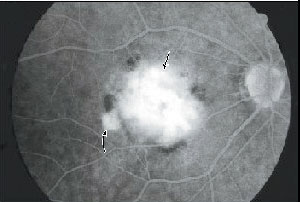 |
| Lesion type and other factors to be assessed for PDT treatment have been updated. |
Criteria include lesion size for AMD patients with either a minimally classic lesion composition (where treatment usually should be considered only for relatively smaller lesions) or occult with no classic lesions (where treatment usually should be considered for relatively smaller lesions or those >4 MPS disc areas with a relatively lower or poorer best-corrected visual acuity) but not patient age, history of systemic arterial hypertension, or prior laser photocoagulation. Therapy should be initiated ideally within one week of the initial fluorescein angiogram on which the clinical decision to treat is based.
Follow-up should be at least every three months (± two weeks) after any initial or subsequent treatment to determine if there is fluorescein leakage from CNV. Additional courses of treatment should be considered as often as every three months (± two weeks) if such leakage is noted at that time. Additional courses of treatment could be deferred if the biomicroscopic and fluorescein angiographic appearances of the lesion are unchanged and show minimal fluorescein leakage, especially when there is no subretinal fluid or fluorescein leakage from CNV underlying the center of the foveal avascular zone.
Patients should avoid exposure of skin or eyes to direct sunlight or bright indoor light for 48 hours after treatment or until resolution of any swelling or discoloration from extravasation. Follow-up of relatively larger minimally classic lesions and occult with no classic lesions that initially do not undergo therapy appears indicated so therapy can be considered if a predominantly classic lesion develops or, in the case of occult with no classic lesions, if visual acuity declines slightly to a lower (poorer) level without a marked increase in lesion size. Additional revisions of these guidelines may be required as new data become available.
(Retina 2005;25(2):119-134)
Verteporfin Roundtable Participants
SLO and Angiography with Wide-Field Contact Lens
Researchers in Italy and the United States performed fluorescein and indocyanine green angiography for large or peripheral chorioretinal structures using a contact lens system that provides a fivefold increase in the field of view of a confocal scanning laser ophthalmoscope.
Separate handheld contact and noncontact ophthalmoscopic lenses were manually aligned with the optical axis of a confocal SLO to demonstrate the feasibility of wide-field SLO angiography. An integrated, wide-field contact lens system was then designed and constructed to increase the SLO's 10-, 20, and 30-degree imaging fields to 50-, 100-, and 150-degree respectively.
Simultaneous fluorescein and ICG angiography was performed with the integrated, wide-field contact lens system for more than 50 patients with disorders that affect their peripheral retina and choroid. Retinal and choroidal abnormalities, including neovascularization and capillary nonperfusion, are easily detected and documented well beyond the range of conventional fundus cameras and SLOs. Peripheral retinal and choroidal hemodynamics can be readily observed and recorded.
The group concludes that confocal SLO has adequate resolution for clinically useful reflectance and angiographic imaging even when its field size is increased fivefold by a wide-field contact lens system. Dynamic and static wide-field angiography can be performed without the limitations of manual or computer-automated photomontages. Peripheral retinal conditions can be studied and recorded to confirm observations from indirect ophthalmoscopy and to facilitate retinal photocoagulation and vitreoretinal surgery.
(Arch Ophthalmol 2005;123:244-252)
Staurenghi G, MD, Viola F, Mainster M, Graham R, Harrington P.


