The appropriate use of antibiotics to prevent infection continues to be a topic of great debate in ophthalmology. In our specialty, there is increasing concern over reports of significant bacterial resistance with current antibiotics. At the same time, there appears to be very little, if any, major decrease in the rate of end-ophthalmitis after various procedures when antibiotics are utilized.
Due to these issues and similar concerns in other specialties, the U.S. Centers for Disease Control and Prevention announced it will be launching a new surveillance system to track antibiotic use in hospitals.1 The CDC has provided funding to four health departments working with academic institutions to establish the new tracking system in 70 hospitals. The new system will automatically transfer drug administration data and bar code records into the tracking system. This will allow providers to compare their antibiotic use with that of other facilities and provide data to promote more judicious use of antibiotics.
In this article, recent endophthalmitis publications will be reviewed that play a significant role in the way we address endophthalmitis, both in terms of prophylaxis as well as current policies and outcomes.
Peri-procedural Prophylaxis
|
A panel of vitreoretinal medical and surgical experts met in 2004 and established a set of guidelines for the preparation, administration and post-procedural management of intravitreal injections based on the available data and their collective experience.4 Of note, no consensus could be reached regarding the value or the need for topical antibiotics before, during or after an intravitreal injection. Generally agreed upon recommendations included the application of povidone-iodine (PI) to the eyelid margin, eyelashes and conjunctival ocular surface prior to injection, and the use of a lid speculum. A number of issues have come to light since the publication of these guidelines. A recent editorial points out the important distinction between topical antiseptic agents compared to commonly used topical antibiotics.5 The use or non-use of face masks is another controversial issue.
|
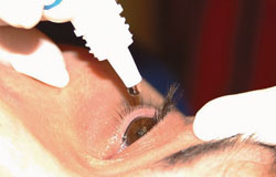
• Topical antibiotics. The use of prophylactic topical antibiotics as a strategy to prevent endophthalmitis is controversial (See Figure 3). Prospective studies have confirmed that topical antibiotics administered one hour before intravitreal injection significantly reduce conjunctival bacteria flora,8,9 and in vitro studies using fourth-generation fluoroquinolones demonstrate eradication of some endopthalmitis-causative organisms in five to 15 minutes.10 Therefore, topical antibiotics would seem to have benefit given the presumed mechanisms of post-injection endophthalmitis, which involve direct inoculation of ocular flora at the time of injection or subsequent entry through a wound track.11 However, topical antibiotics have demonstrated no additional effect on reducing conjunctival bacterial counts beyond the effect of 5% PI alone.12,13
A recent randomized, controlled, longitudinal study demonstrated that repeated exposure of both ocular and nasopharyngeal flora to ophthalmic antibiotics as prophylaxis for intravitreal injections selects for resistant strains of bacteria.14 The results of this study showed macrolide- and fluoroquinolone-resistant conjunctival coagulase-negative staphylococci emerging rapidly after exposure to their respective antibiotic. Of key significance, the resistance was maintained or worsened by periodic reexposure. This finding is thought to have considerable implications because conjunctival flora are presumed to be the predominant source of post-injection endophthalmitis, and because antibiotic-resistant S. epidermidis is associated with greater intraocular inflammation, virulence and increased ocular infection rate compared to antibiotic-susceptible strains.15,16 A study we conducted further demonstrates the alarming increase in fluoroquinolone resistance among coagulase-negative staphylococcus endophthalmitis isolates, with rates of resistance of each of the fluoroquinolone antibiotics at more than 50 percent (in press, Archives of Ophthalmology).
|
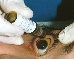
• Intracameral antibiotic agents for cataract surgery. A multicenter, prospective, randomized study conducted by the European Society of Cataract and Refractive Surgeons provided positive data supporting the use of a direct intracameral bolus of cefuroxime at the conclusion of cataract surgery (See Figure 4).19 A recent update to this study demonstrates seven-year follow-up data of the study and continued support of this practice.20 The study initially demonstrated rates of culture-proven infectious endophthalmitis at 0.07 percent in the groups receiving intracameral cefuroxime prophylaxis compared with rates of 0.34 percent in the control groups not receiving intracameral prophylaxis. Despite this significant difference, concerns immediately surfaced because the control group (operated on from January 1996 through December 2002) had such an elevated rate of endophthalmitis compared to other studies performed during the same time period (0.04 percent,21 0.128 percent22). Rather than settling the debate, the ESCRS fueled further controversy on the choice of antibiotic and value of this practice.
Cefuroxime is a second-generation cephalosporin with antibacterial activity resulting from its ability to inhibit bacterial cell wall synthesis. It has limited efficacy against gram-negative bacteria and high rates of resistance against methicillin-resistant Staphylococcus epidermidis and Staph. aureus, two of the most common pathogens in post-cataract surgery endophthalmitis infections. Cefuroxime is not available in a prepackaged form for intracameral use and must be diluted from powder form in the operating room, yielding significant concerns for dilution errors and contamination.23 Cases of dilutional errors with intracameral antibiotics, including cefuroxime and vancomycin, have reportedly resulted in macular thickening, chronic cystoid macular edema, serous retinal detachment and macular infarction. Concerns of intracameral antibiotics causing toxic anterior segment syndrome (TASS), an acute, severe, sterile postoperative inflammation, have also limited the regular use of intracameral antibiotics in the United States.24 Their effectiveness is further questioned, as the safe concentration of antibiotics in the irrigation fluid requires more than 140 minutes to exhibit a bactericidal effect, and it is reported that the half-life of antibiotics in the anterior chamber is only 51 minutes.25,26
Since contemporary endophthalmitis rates after cataract surgery without intracameral antibiotics (0.03 percent27) are equivalent to or lower than the ESCRS study rates with intracameral antibiotics (0.05 percent20), there appears to be little support for their use given the significant risks.
The Role of Face Masks
Two large studies have been recently released demonstrating a significantly elevated percentage of streptococcal species involved in post-injection endophthalmitis isolates compared to postsurgical end-ophthalmitis isolates. The first study was a large meta-analysis of end-
ophthalmitis after intravitreal injection of anti-VEGF agents including most major U.S.-based studies from 2005 to 2010.2 In this evaluation, streptococcal isolates were approximately three times more frequent after intravitreal anti-vascular endothelial growth factor injection than after intraocular surgery.
Another group conducted a study over a similar six-year time period including 60,322 patients from a single institution undergoing intravitreal injection and reported that five of seven culture-positive cases were due to streptococcal species.3 The result of these studies raised concerns regarding the possible oropharyngeal origin of these streptococcal species. The author of the first study proposed strategies to consider minimizing oropharyngeal moisture droplet transmission, including avoiding talking, coughing or sneezing, and wearing surgical face masks during injection procedures. If a true attempt to reduce aerosolized moisture droplets is recommended, then the physician, the nurse/assistant and the patient should all wear face masks.
As a follow-up to these studies, a simulation of injection conditions was created to demonstrate the potential oropharyngeal droplet contamination of the field during intravitreal injection.28 The authors concluded that significantly more colony-forming bacteria are dispersed onto an agar plate when speaking without a face mask compared with when wearing a face mask or remaining silent during a simulated intravitreal injection procedure. However, when povidone-iodine was utilized on the agar plates prior to a similar in vitro procedure, the dispersed oral flora were effectively eliminated, suggesting that the use of face masks is less necessary with adequate antiseptic measures.29
|
Compounding Anti-VEGF Meds
In South Florida, a recent outbreak of post-injection endophthalmitis in 12 patients was reported after intravitreal injection of bevacizumab (Avastin).31 All patients received topical antibiotics after the intravitreal injection. All patients received intravitreal injections of bevacizumab prepared by the same compounding pharmacy in South Florida and 10 of 12 patients had positive microbiologic cultures for Streptococcus mitis/oralis. Seven unused syringes of bevacizumab prepared by the same compounding pharmacy at the same time also were also positive for S. mitis/oralis. The visual outcomes were poor in these cases, consistent with previous studies reporting poor visual outcomes in streptococcal endophthalmitis.32
While the majority of the literature on reducing endophthalmitis rates after intravitreal injection has focused on the peri-injection period, the importance of drug preparation has largely been ignored. Bevacizumab is distributed in 4- or 16-mL, preservative-free, single-use vials, and is typically aliquoted into smaller doses for intravitreal use. The report describes the importance of compounding pharmacist compliance with the standards outlined in United States Pharmacopeia chapter 797. These measures include the use of trained and certified staff, personal protective equipment and a properly operated and certified ISO class 5 environment. The study additionally calls into question whether bevacizumab syringes prepared from two different batches might be preferred in patients requiring bilateral injections.
The use of peri-procedural topical antibiotics when povidone-iodine is utilized may not reduce endophthalmitis rates, imposes a large monetary burden on our health-care system, and is likely contributing to the rise in bacterial resistance, particularly after repeated fluoroquinolone usage. Physicians performing intravitreal injection procedures are now aware of the relatively increased frequency of post-injection streptococcal endophthalmitis. Precautions include minimizing speech during the procedure and adherence to an aseptic protocol. Strict pharmacist compliance with USP chapter 797 mandates is important in the preparation of bevacizumab in order to reduce the chance of future outbreaks of post-intravitreal injection endophthalmitis. In general, cataract surgeons have not adopted the use of intracameral antibiotics and yet the rates of endophthalmitis remain very low. REVIEW
Dr. Schimel is a fellow in vitreoretinal surgery and a clinical instructor at the Bascom Palmer Eye Institute. Dr. Flynn is the head of the Retina Service and the J. Donald M. Gass Distinguished Chair at the Bascom Palmer Eye Institute at the University of Miami Miller School of Medicine, Miami. Contact Dr. Flynn at:
hflynn@med.miami.edu. This work was supported in part by the National Institute of Health Center (grant P30-EY014801) and an unrestricted grant from Research to Prevent Blindness, New York. The authors have no financial or proprietary interest in the materials presented herein.
1. Kuehn B. Antibiotic Use Tracking. JAMA 2011;306(24):2661-2661.
2. McCannel CA. Meta-analysis of endophthalmitis after intravitreal injection of anti-vascular endothelial growth factor agents: Causative organisms and possible prevention strategies. Retina 2011;31:654-661.
3. Moshfeghi AA, Rosenfeld PJ, Flynn HW Jr., et al. Endoph-thalmitis after intravitreal anti-vascular endothelial growth factor antagonists: A six-year experience at a university referral center. Retina 2011;31:662-668.
4. Aiello LP, Brucker AJ, Chang S, et al. Evolving guidelines for intravitreous injections. Retina 2004;24(5 Suppl):S3-19.
5. Wykoff CC, Flynn HW, Jr., Rosenfeld PJ. Prophylaxis for end-ophthalmitis following intravitreal injection: Antisepsis and anti-biotics. Am J Ophthalmol 2011;152:717-719 e712.
6. Berkelman RL, Holland BW, Anderson RL. Increased bactericidal activity of dilute preparations of povidone-iodine solutions. J Clin Microbiol 1982;15:635-639.
7. Hosseini H, Ashraf MJ, Saleh M, et al. Effect of povidone-iodine concentration and exposure time on bacteria isolated from endophthalmitis cases. J Cataract Refract Surg 2012;38:92-96.
8. Ta CN, Egbert PR, Singh K, Shriver EM, et al. Prospective randomized comparison of 3-day versus 1-hour preoperative ofloxacin prophylaxis for cataract surgery. Ophthalmology 2002;109:2036-2040; discussion 2040-2031.
9. Moss JM, Nguyen D, Liu YI, et al. Comparison of one-day versus one-hour application of topical gatifloxacin in eliminating conjunctival bacterial flora. Ophthalmology 2008;115:2013-2016.
10. Callegan MC, Novosad BD, Ramadan RT, Wiskur B, Moyer AL. Rate of bacterial eradication by ophthalmic solutions of fourth-generation fluoroquinolones. Adv Ther 2009;26:447-454.
11. Kim SJ, Toma HS, Midha NK, Cherney EF, et al. Antibiotic resistance of conjunctiva and nasopharynx evaluation study: A prospective study of patients undergoing intravitreal injections. Ophthalmology 2010;117:2372-2378.
12. Moss JM, Sanislo SR, Ta CN. A prospective randomized evaluation of topical gatifloxacin on conjunctival flora in patients undergoing intravitreal injections. Ophthalmology 2009;116:1498-1501.
13. Halachmi-Eyal O, Lang Y, Keness Y, Miron D. Preoperative topical moxifloxacin 0.5% and povidone-iodine 5.0% versus povidone-iodine 5.0% alone to reduce bacterial colonization in the conjunctival sac. J Cataract Refract Surg 2009;35:2109-2114.
14. Kim SJ, Toma HS. Antimicrobial resistance and ophthalmic antibiotics: 1-year results of a longitudinal controlled study of patients undergoing intravitreal injections. Arch Ophthalmol 2011;129:1180-1188.
15. Mino De Kaspar H, Hoepfner AS, Engelbert M, et al. Antibiotic resistance pattern and visual outcome in experimentally-induced Staphylococcus epidermidis endophthalmitis in a rabbit model. Ophthalmology 2001;108:470-478.
16. Miller D, Flynn PM, Scott IU, Alfonso EC, Flynn HW, Jr. In vitro fluoroquinolone resistance in staphylococcal endophthalmitis isolates. Arch Ophthalmol 2006;124:479-483.
17. Hyon JY, Eser I, O’Brien TP. Kill rates of preserved and preservative-free topical 8-methoxy fluoroquinolones against various strains of Staphylococcus. J Cataract Refract Surg 2009;35:1609-1613.
18. Costello P, Bakri SJ, Beer PM, et al. Vitreous penetration of topical moxifloxacin and gatifloxacin in humans. Retina 2006;26:191-195.
19. Endophthalmitis Study Group, ESCRS. Prophylaxis of postoperative endophthalmitis following cataract surgery: Results of the ESCRS multicenter study and identification of risk factors. J Cataract Refract Surg 2007;33:978-988.
20. Romero-Aroca P, Mendez-Marin I, Salvat-Serra M, Fernandez-Ballart J, et al. Results at seven years after the use of Intracamerular cefazolin as an endophthalmitis prophylaxis in cataract surgery. BMC Ophthalmol 2012;12:2.
21. Eifrig CW, Flynn HW Jr., Scott IU, Newton J. Acute-onset postoperative endophthalmitis: review of incidence and visual outcomes (1995-2001). Ophthalmic Surg Lasers Sep-Oct 2002;33(5):373-378.
22. Taban M, Behrens A, Newcomb RL, et al. Acute endophthalmitis following cataract surgery: A systematic review of the literature. Arch Ophthalmol 2005;123:613-620.
23. Montan PG, Wejde G, Setterquist H, Rylander M, et al. Prophylactic intracameral cefuroxime. Evaluation of safety and kinetics in cataract surgery. J Cataract Refract Surg 2002;28:982-987.
24. Mamalis N, Edelhauser HF, Dawson DG, Chew J, et al. Toxic anterior segment syndrome. J Cataract Refract Surg 2006;32:324-333.
25. Gritz DC, Cevallos AV, Smolin G, Whitcher JP, Jr. Antibiotic supplementation of intraocular irrigating solutions. An in vitro model of antibacterial action. Ophthalmology 1996;103:1204-1208; discussion 1208-1209.
26. Lehmann OJ, Thompson JP, White LO, Keys MF, Campbell MJ. Half-life of intracameral gentamicin after phacoemulsification. J Cataract Refract Surg 1997;23:883-888.
27. Wykoff CC, Parrott MB, Flynn HW, Jr., Shi W, Miller D, Alfonso EC. Nosocomial acute-onset postoperative endophthalmitis at a university teaching hospital (2002-2009). Am J Ophthalmol 2011;150:392-398 e392.
28. Wen JC, McCannel CA, Mochon AB, Garner OB. Bacterial dispersal associated with speech in the setting of intravitreous injections. Arch Ophthalmol 2011;129:1551-1554.
29. Doshi RR, Leng T, Fung AE. Reducing Oral Flora Contamination of Intravitreal Injections with Face Mask or Silence. Retina 2012;32:473-6.
30. Schimel AM, Scott IU, Flynn HW, Jr. Endophthalmitis after intravitreal injections: Should the use of face masks be the standard of care? Arch Ophthalmol 2011;129:1607-1609.
31. Goldberg RA, Flynn HW Jr, Isom RF, Miller D, Gonzalez S. An Outbreak of Streptococcus Endophthalmitis After Intravitreal Injection of Bevacizumab. Am J Ophthalmol 2012;153:204-208.e1.
32. Miller JJ, Scott IU, Flynn HW Jr., Smiddy WE, et al. Endoph-thalmitis caused by Streptococcus pneumoniae. Am J Ophthalmol 2004;138:231-236.


