Wavetec Vision last month launched its ORange Intraoperative Wavefront Aberrometer. So-named because it is used in the "OR" on a "range" of applications, the aberrometer attaches to the surgical microscope and provides on-demand readings of sphere, cylinder and axis in approximately two to five seconds. The system also has a large dynamic range, -5 D to +20 D, making it ideal for intraoperative measurements.
With
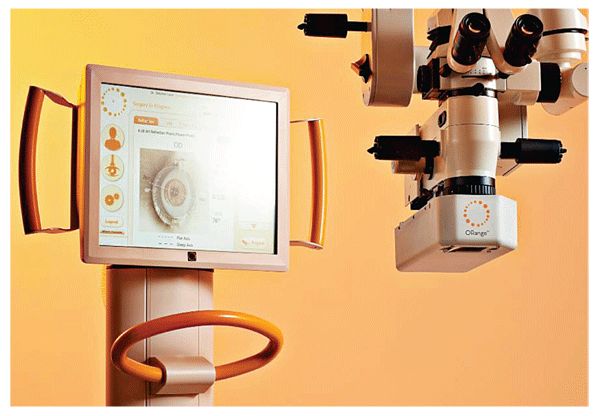
ORange uses an interferometer to analyze the wavefront reflected out of the eye by relaying it through an optical system and directing it through a pair of gratings that are set at a specific distance from, and offset angle to, each other. This creates a diffraction, or fringe, pattern. The gratings are fixed at an optimal angle with respect to one another, which creates a high-resolution optical effect. This fringe pattern is captured by a camera and then processed using Wavetec Vision proprietary algorithms to create the measurement. Each fringe pattern will be unique for every eye.
Initial applications will be for guiding LRIs and ensuring proper axis placement of toric IOLs. A future application will be the use of the
Surgeons will have information regarding the optical system of the eye at the click of a footswitch, data that has not been available to them previously during the operative procedure. Knowing the refraction of the eye after various steps in the surgery will help guide their decision-making.
The aberrometer will also capture all of the data gathered intraoperatively and transfer it automatically to the ORange AnalyzOR database, giving surgeons the ability to track and compare data via individualized reports.
The clinical evaluation of the ORange Intraoperative Wavefront Aberrometer has been under way at eight sites around the
New Solutions for Visually Impaired
Optelec
Optelec FarView is a powerful and portable magnification solution designed for the active lifestyle that redefines how low-vision users are able to access, store, review and share information in an ultra compact and stylish design. It includes a built-in auto focus camera, zoom-in functionality, scrolling and auto-return allow the user to bring the environment within the scope of vision or preferred retinal locus for best visual acuity.
Optelec ClearView+ G2 is a desktop electronic video magnifier that allows the user to read and write with magnified and illuminated viewing assistance. Its five-directional arm with 90-degree rotating screen and pull flexibility make it easy to maneuver for the low-vision user. HD image quality provides high resolution and smooth de-interlaced images to increase the user's reading time and pleasure by eliminating image fading and eye fatigue. Optelec ClearNote Portable is a lightweight video magnifier that combines distance, document or self-viewing modes. The ClearNote Portable connects to any desktop or laptop computer via the USB port. Call 1 (800) 335-7970 or visit optelec.com.
TrueVision Adds 3D Flat Panel Displays
TrueVision Systems has introduced new flat panel product configurations enabling operating rooms and exam rooms to have 3D 1080p flat panel LCDs to display live and recorded content of the surgical view from a microscope or slit lamp.
Different configurations include wall mounted and portable ergonomic cart solutions with a selection of high quality LCD 3D 1080p panels ranging from 24 to 46 inches. One or more displays can be mounted on the wall, from the ceiling, or on a boom arm for operating rooms, exam rooms, and offices.
TrueVision says its 3D visualization system for microsurgery enables surgeons to perform or view surgery via a heads-up display instead of looking through the microscope. It features the ability to record, edit and playback 3D 1080p operative content. The system is designed to seamlessly bring patient images and data from the exam room into the OR.
All systems include the patented TrueVision 3D Image Capture Module that attaches to the surgical microscope or slit lamp, an Image Processing and Recording Unit and choice of a display from 24 inches to 46 inches on a cart or wall/ceiling mounted. The TrueVision Surgical Vision System is registered with the FDA as a class one medical device. For information, call (805) 963-9700 or visit truevisionsys.com.
Patient-Friendly Tonometer with Silent and Soft Air Pulse
Tomey
PEARLS FROM THE DEEP
A Two-Step Avoids Collapse and Excessive Deepening of the AC
There is increasing awareness that a stable anterior chamber depth avoids trampolining of the vitreous body and may reduce the incidence of pseudophakic retinal detachment.
Chamber collapse is common not only in distensible myopic eyes but in shallow-chambered hyperopic eyes. Maintenance of positive pressure is important to discourage posterior fluid misdirection syndrome and choroidal effusion or hemorrhage in very short eyes. The chamber is most likely to shallow upon removal of the phaco tip or the irrigation and aspiration (I&A) tip from the eye. We have all learned to place viscoelastic through the side port when there is a broken capsule before withdrawal of the phaco tip to avoid vitreous prolapse. I believe we should apply this principle to routine cases.
Therefore, I employ two maneuvers which discourage anterior chamber collapse and over-deepening.
Although a chamber maintainer is effective, it requires a second source of irrigation and an extra incision. Instead, at the conclusion of phaco, I trade my chopper for BSS in a syringe with cannula through the side port prior to withdrawal of the primary instrument in all routine cases. I can then irrigate manually as I withdraw the phaco or I&A tip, allowing the internal Descemet's valve to seal, thereby maintaining positive pressure in the few seconds it takes to get the next instrument. The syringe is then also handy to irrigate the incision, clearing out debris if it does not spontaneously seal. I can use the cannula through the side port for fixation and be prepared to finally seal the incision at the conclusion of the case without risking anterior chamber collapse with lens-endothelial touch.
The second maneuver avoids over-deepening of the anterior chamber, most noticeable in the detachment-prone myope. The reason for extreme deepening of the chamber has been postulated to be a reverse pupillary block mechanism where the pupil edge contacts the anterior capsule flap for 360 degrees, preventing fluid from distributing between the anterior and posterior chambers. In the at-risk eye (pars plana vitrectomy, loose zonules or myope) I will place a bolus of viscoelastic between the iris and the capsule in 1 or 2 clock hours just after hydrodissection and before entry with the phaco tip. I will also lower the bottle upon initial entry so the rush of fluid into the anterior chamber is less forceful. As soon as equilibrium is established, I raise the bottle to its normal level. This will usually prevent the reverse block. If the OVD isn't effective in preventing reverse block, then the tip of the non-dominant hand instrument is insinuated between the pupil edge and the anterior capsule. As the iris is lifted away from the surface of the capsule, the chamber will shallow to a more appropriate depth. Rarely, this can be segmental and may need to be lifted in more than one quadrant.
Be mindful of maintaining homeostatic chamber depth to lessen the influx of surface debris, discourage misdirection of fluids and prevent stress on the ciliary body and vitreous base. The described maneuvers lead to more comfortable patients intraoperatively; quieter eyes perioperatively and, possibly, fewer long term retinal sequelae.


