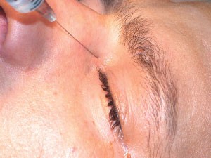
Physicians at the Gülhane Military Medical Academy in Istanbul, Turkey, collected data over an eight-year period (1996-2004) on young patients (mean age: 22.64 ±1.71) undergoing external DCR at the Istanbul Academy. The patients were randomly separated into two groups: general anesthesia and local anesthesia. Of the 480 DCR procedures, 182 were performed under general anesthesia (44 bilateral) and 298 were performed using local anesthesia (32 bilateral). In the two-hour postoperative period, postop nausea and vomiting, epistaxis, length of hospital stay, and intraoperative bleeding were documented.

From the results of the study the physicians conclude that administering local anesthesia in young patients undergoing external DCR is a good alternative because it is cost-effective and eliminates the complications associated with the administration of general anesthesia.
(Ophthal Plast Reconstr Surg 2005;21(3)201-205)
Ciftci F, Pocan S, Karadayi K, Gulecek O
Self-Sealing Corneal Tunnel Incision Deemed Safe
CLEAR CORNEA CATARACT SURGERY PERFORMED UNDER topical anesthesia is safe and results in few postoperative complications, according to a nine-year study by two ophthalmologists in New Orleans.
One surgeon performed the topical clear cataract surgery using a self-sealing corneal tunnel incision for more than nine years and had previously reported success with the procedure. A total of 3,500 consecutive clear cornea topical anesthesia cataract surgeries were performed from January 1994 until May 2003. Only consecutive cases of clear cornea cataract surgery were included in the study. All cases were operated using phacoemulsification via a temporal 3 x 2-mm clear cornea incision with lidocaine hydrochloride gel (20 mg/ml) applied over the eye in three doses every 10 minutes for 30 minutes before surgery. Of the 3,500 surgeries performed, 56 (1.6 percent) required a suture from wound leakage occurring before completion of the surgery. None of the 3,500 cases required a return to the operating room, and six cases (0.17 percent) had a retinal detachment within 30 days postop.
(Ophthalmology 2005; 112:985-986)
Monica M, Long D.
Glaucoma Noncompliance
NONCOMPLIANCE WITH HYPOSENSITIVE TREATMENT was found to be common among glaucoma patients, according to a review of scientific literature studies conducted in the Netherlands. However, no strong evidence was found to support a relationship between noncompliance and progressive visual field loss.
Physicians at the Department of Epidemiology and the Department of Ophthalmology, Maastricht, the Netherlands, reviewed the literature in MEDLINE, EMBASE, CIN-AHL, PsychInfo, and Cochrane databases. They included 34 articles de-scribing 29 quantitative studies in English, German, French or Dutch, excluding studies on noncompliance in drug trials.
Their review found that the proportion of patients who deviate from prescribed medication therapy regimen extended from 5 to 80 percent. The effect of noncompliance on clinical outcome has not yet been determined, and no factors sensitive and specific enough have been realized in identifying potential noncompliant patients precisely. However, identifiable factors in improving compliance include patient knowledge and dose frequency. Therefore, combining patient education and preclusion of forgetting doses may assist in enhancing successful patient compliance.
(Ophthalmology 2005; 112:985-986)
Olthoff C, Schouten J, van de Borne B, Webers C.
LASIK Effective Following Phacoemulsification
UNDERGOING LASIK FOLLOWING CATARACT SURGERY appears to be effective in correcting refractive errors, according to an Australian study.
Twenty-three eyes of nineteen patients (10 female, nine male) were reviewed at the Eye Institute, Sydney, after being treated with LASIK for refractive error following cataract surgery. The Summit Apex Plus and LADARVision excimer laser and the SKBM microkeratome were used. The mean age of the patients was 63.5 years (range 50 to 88), and the mean length of follow-up time was 8.4 months (range, one to 12 months). The mean interval between the pa-tient undergoing cataract surgery and LASIK was 12 months (range 2.5 to 46 months.)
The mean preoperative spherical equivalent refraction for myopic eyes was
-3.08 ±0.84 D (range -4.75 to –2D). For hyperopic eyes it was +1.82 ±1.03 D (range +0.75 to +3D). The mean improvement following LASIK surgery was greater for myopic than hyperopic eyes (myopic, 2.54 ±1.03 D versus hyperopic, 1.73 ±0.62 D; P=.033). The percentage of eyes with uncorrected visual acuity of 20/40 or better in the myopic group im-proved from none preoperatively to 91.7 percent postop (P<.001). The patients in the hyperopic group showed improvement from 27.3 percent preop to 90.9 percent postop (P<.008). No eyes lost two or more lines of best-corrected visual acuity.
Though LASIK seems to correct refractive error after cataract surgery, more long-term studies are necessary to determine refractive stability.
(J Cataract Refract Surg 2005; 31:979-986)
Kim P, Briganti E, Sutton G, Lawless M. Rogers C, Hodge C.
Intacs Segments Effective for Keratoconus Correction
ONE OR TWO INTACS SEGMENTS ORIENTED BY A PREOPERATIVE corneal topography pattern proved to be effective in decreasing the corneal steepening and astigmatism in the treatment of keratoconus, according to a Spanish study. Improved best-corrected visual acuity was also noted.
The study was conducted at Miguel Hernandez University, Alicante, on 26 keratoconic eyes with clear central corneas of 19 consecutive patients (nine women and 10 men). According to the topographic pattern of the cone, corneas were separated into two groups: Group I comprised keratoconus not crossing the 180º meridian; Group II comprised keratoconus crossing the 180º meridian. The Intacs segments were positioned horizontally through a lateral clear corneal incision. Following the corneal topography, Group I received one segment implanted 0.45 mm inferior. Group II had two segments implanted—one 0.25 mm superior and one, 0.45 mm inferior. All patients completed a minimum follow-up of one year.
Results showed significant reduction in spherical equivalent error and refractive astigmatism. For both groups, the mean keratometric values were reduced following Intacs insertion. After one year of follow-up, Group I (receiving one segment) had an improvement in mean uncorrected visual acuity to 20/50 (0.4 ±0.22 decimal value). This was a statistically significant finding when compared to the preop uncorrected visual acuity of 20/100 (0.2 ±0.13 decimal value) (P=.011).
The study recommended further follow-up to determine the final effect of Intacs on the progression of keratoconus.
(J Cataract Refract Surg 2005; 31:943-953)
Alió J, Artola A, Hassanein A, Haroun H, Galal A.
Intravitreal Triamcinolone Injection Induces Cataracts
SINGLE INTRAVITREAL TRIAMCINOLONE INJECTION induces posterior subcapsular cataract development, while multiple injections result in all-layer cataract progression, according to a New York study. Local administration of triamcinolone causes high corticosteroid concentrations near the designated tissues, and though minimal systemic effects occur, intravitreal corticosteroids do induce local adverse affects.
For the study, physicians injected intravitreal triamcinolone in 42 phakic eyes of 37 patients. Each patient was injected one, two or three times for a variety of indications. The patients' non-injected phakic eyes served as the control. Follow-up times were 12 months for single injection, 14 months for multiple injections, and 13 months for the controls.
Changes in posterior subcapsular cataract and refractive error from baseline proved significantly different between single intravitreal triamcinolone injected eyes and the control group [0.7 ±0.2 (mean ±SEM [arbitrary unit] vs. 0.2 ±0.1, P=.02; and -0.5 ±0.1 D vs -0.2 ±0.1 D, P=.01, respectively) at the last follow-up visit. For multiple-injected eyes and control eyes change from baseline in corticonuclear cataract (1.1 ±0.2 vs. 0.2 ±0.1), posterior subcapsular cataract (1.1 ±0.2) and refractive error (-1.8 ±0.4 D) were significantly different (P<.001, P<.001, and P<.001, respectively).
(Am J Ophthalmol 2005;139:993-998)
Çekiç O, Chang S, Tseng J, Akar Y, Barile G, Schiff


