The threat of ectasia hangs over every laser refractive procedure, and many hours are spent trying to develop new and better ways to detect patients at risk for it preop in order to avoid operating on them. In this article, diagnosticians explain their preferred methods to screen patients for suspicious corneal findings preop and to detect the beginnings of ectasia postop.
Emphasizing Elevation
Michael Belin, MD, a corneal specialist and refractive surgeon from
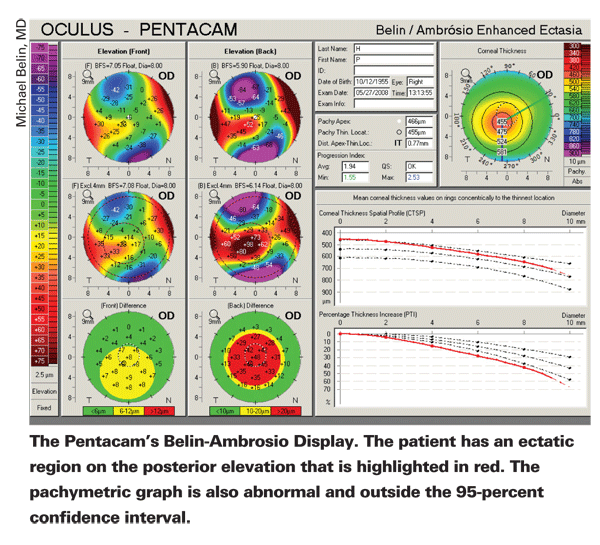
"In the past, many clinicians have tried to diagnose keratoconus just by looking at the anterior surface of the cornea," says Dr. Belin. "But, by definition, keratoconus or ectasia is a corneal thinning, and this involves two surfaces, the anterior and the posterior. By ignoring the posterior surface, and by ignoring the pachymetric or corneal thickness distribution, you're probably ignoring the earliest, most sensitive and most specific signs of keratoconus and ectasia. So, I'm a proponent of elevation maps because they display information from both the anterior and the posterior surfaces. And, if you have accurate anterior/posterior information you then have the corneal thickness profiles, or the pachymetric maps. On almost every keratoconus patient I've seen, the changes on the posterior surface are more prominent and sometimes occur in the absence of any changes on the anterior surface."
In an effort to put elevation data to use, Dr. Belin has teamed with Brazilian surgeon Renato Ambrosio Jr. to create a special screening display for the Oculus Pentacam. Dr. Belin is a consultant to Oculus.
The color-coded display shows normal maps as green and posterior-surface maps that are three standard deviations from normal as red. Yellow areas aren't necessarily abnormal, but are there to show the gradations between the two opposite ends. "It's basically a display that further highlights the areas of keratoconus," says Dr. Belin. "What we did was make a reference surface that more closely resembles the normal, non-conical portion of the cornea. This, then, highlights a cone even more. We combined this with Dr. Ambrosio's pachymetric indices and graphs to get a display that only has elevation data—since pachymetry is really elevation data—that we can use to screen patients for ectasia."
The reference surface that's used is an important part of any screening process, since it sets what the computer views as "normal." To get normal values and keratoconic counterparts, Drs. Belin and Ambrosio input the corneas of 800 patients, some normal, some keratoconic. Dr. Belin says they're currently amassing more patient data to expand this database. "We use a best-fit sphere with a reference surface diameter of 8 mm," he explains. "We found this diameter fits to the more normal portion of the cornea without getting into the limbal region, and ensures that, most of the time, you're getting accurate, non-extrapolated data. Under these conditions, the average anterior elevation of the corneal apex for normal patients was 1.6 ±1.3 µm. For the thinnest point it was 1.7 ±2 µm. On the posterior surface of normal corneas, the apex was 0.8 ±3 µm and the thinnest point was 3.6 ±4.7 µm. At two standard deviations from normal, it was 4.2 µm for the apex and 5.5 µm for the thinnest point of the anterior cornea, and 6.8 µm for the apex and 9.8 µm for the thinnest point for the posterior." Three standard deviations away from normal on the posterior surface, which is used for screening, is 13 µm for the apex and 17.7 µm for the thinnest point.
Catching Ectasia Postop
Ophthalmic researcher Stephen D. Klyce, PhD, of the Department of Ophthalmology, Mt Sinai School of Medicine in New York City, says the signs of ectasia can be subtle initially and could possibly be mistaken for contact lens warpage, the effects of trauma or a normal, though undesirable, healing event. Conversely, the patient may not have ectasia, but a topographer's dioptric scale makes it appear that he does. Here are his suggestions for detecting true ectasia.
"The etiology of ectasia is fairly well understood. It can have pathologic or iatrogenic underpinnings or it can result from a combination of both in rare cases when refractive surgery has exacerbated corneal pathology," says Dr. Klyce. "In pathologic ectasia, there is a pre-existing pathology such as keratoconus or pellucid marginal degeneration. These are both stromal thinning diseases that can biomechanically result in corneal bulging. Both pathologies are contraindications for refractive surgery as further weakening of the cornea increases the risk for the development of ectasia. It can be argued that keratoconus would have produced ectasia on its own, with or without the refractive procedure, but at the same time, a large percentage of keratoconus patients do stabilize with time.
"The iatrogenic form of ectasia is related to the refractive procedure itself," Dr. Klyce continues. "Some corneas are weaker than others, and if a LASIK flap is too thick or sculpting is too deep, ectasia can develop in the absence of corneal disease."
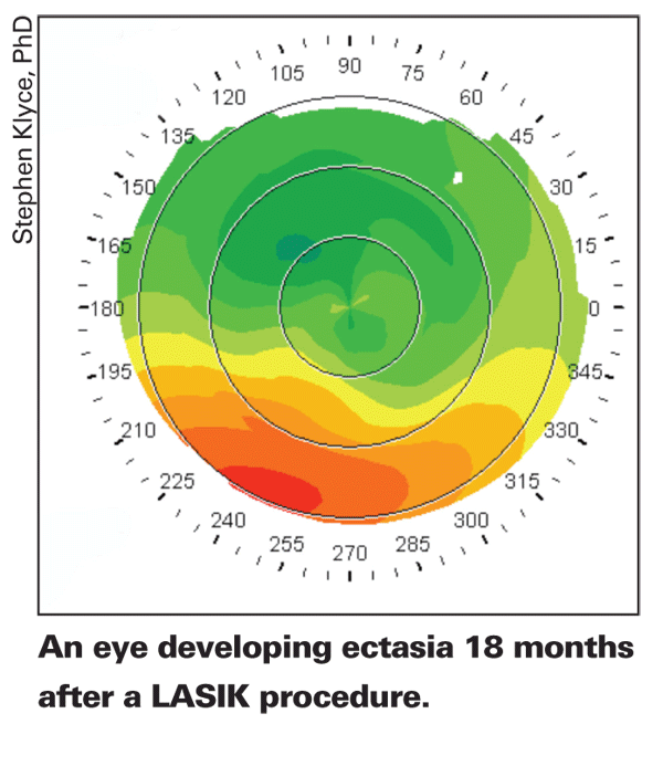
For the patient suspected of having postoperative ectasia, Dr. Klyce recommends collecting all of the corneal topography exams of the patient, preop and postop, including those from any co-managing doctors. When examining these, he stresses the key to understanding changes in corneal topography is to use the same fixed dioptric scale for each one. Often it's necessary to reprint the topography maps after manually adjusting the topographer to use a standard scale. "After LASIK, some topographers will make even stable corneas seem to have a peripheral ectasia by using a scale that displays the peripheral corneal powers in the red region," says Dr. Klyce. He also recommends that, in addition to surveying the available historical topography maps, if the diagnosis isn't obvious, obtain a current corneal topography examination and another in about three months to determine if there are progressive changes that might herald the onset of ectasia. Progressive changes are most easily demonstrated using a topographer's difference map.
Also, if the patient is wearing contact lenses, have these discontinued for about a month and then obtain a repeat topography, says Dr. Klyce, to rule out contact lens warpage.
On the sequential topography maps, Dr. Klyce says the surgeon should note the stability of the topography with the difference map; ectasia presents as a regional progressive increase in power. One common sign is a change in the apparent outline of the ablated area on the power contour map. An inferior bulge is typical and can make the ablation zone smaller in the area of the ectasia. If there's a progressive but uniform increase in power in the ablation zone, this could indicate corneal healing rather than ectasia, resulting from the activation of stromal keratocytes or epithelial hyperplasia. Dr. Klyce also mentions that, like keratoconus, ectasia can develop anywhere on the corneal surface. Says he, "Though ectasia usually appears in the inferior quadrant, there are notable exceptions."


