The Food and Drug Administration has approved Merck’s Zioptan (tafluprost ophthalmic solution) 0.0015%, the first preservative-free prostaglandin analog ophthalmic solution. Zioptan is approved for reducing elevated intraocular pressure in patients with open-angle glaucoma or ocular hypertension.
Zioptan may gradually change eyelashes and vellus hair in the treated eye. These changes include increased length, color, thickness, shape and number of lashes. Eyelash changes are usually reversible upon discontinuation of treatment.
The FDA approval of Zioptan was based on efficacy and safety results from five controlled clinical studies in 905 patients. Both preservative-containing and preservative-free formulations of tafluprost were used in these clinical studies.
In clinical studies of up to two years in duration, Zioptan, dosed once-daily in the evening lowered IOP by 6 to 8 mmHg at three months and by 5 to 8 mmHg at six months, from a baseline pressure of 23 to 26 mmHg. For information, visit
merck.com.
Enhanced Viewing with New Kowa Retinal Camera
Kowa Optimed received 510(k) approval from the FDA for the marketing of its latest model, the VX-20 retinal camera. Like the VX-10, the VX-20 offers non-mydriatic, mydriatic and FA modes with 50-degree and 30-degree views.
Additionally the VX-20 has an autofluorescence mode to provide further diagnostic investigation of the health of the retina. Linked to VK-2 software, the VX-20 provides options for fundus image analysis. With the built-in PC and high resolution camera back, this fundus camera is a versatile diagnostic tool with stand-alone capability.
Kowa VX-20 features include: the optional VK-2 high performance and digital imaging software, which allows for enhanced viewing and image manipulation for further diagnostic investigation; a built-in PC with a high- resolution camera back; and Digiversal software that automatically converges patient image files from multiple pieces of diagnostic equipment into a single file on a single screen. This software solution makes image organization and comparison seamless and easy, the company says. For information, call 1 (800) 966-5692 or visit
kowa-usa.com/eyecare.
 |
 |
Although a thin LASIK flap minimizes the risk of postoperative corneal ectasia, sub-Bowman’s keratomileusis using an automated microkeratome or femtosecond laser can sometimes be associated with folds and microstriae. This may require retreatment and is viewed unfavorably by patients.
|
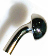
Moria says that’s that’s why it launched a dedicated hand-held instrument for external flap massage that can be used either intraoperatively (preventive massage) and/or in the postoperative time (therapeutic massage); the Caro Manipulator has a hemispherical, highly polished tip to avoid damage to the epithelium.
For more information, visit
moria-surgical.com.
Precision Low-Vision
If you have patients struggling with low vision, Precision Vision has introduced a new line of hand magnifiers that may be able to help.The Ergo-Lux hand magnifiers are illuminated, and feature a four-chip, surface-mounted light-emitting diode. The company says the Ergo-Lux’s design supports the user’s hand and arm to ensure a natural, stress-free reading position, and that the correct distance between the object and the lens is automatically found when the device’s handle sits on the object. Precision Vision adds that the SMD LED generates very bright, consistent, high-contrast light that is the ideal illumination for the visual task at hand. The Ergo-Lux runs on three AAA batteries that the company says are easy to change, thanks to a special closing mechanism in the device’s handle.
The Ergo-Lux comes in five magnification powers between 8 and 24 D in 4-D increments, but is also available as a boxed set with all of the powers. For information, visit
precision-vision.com.
Anterior Segment Module Adds to Spectralis Capabilities
The FDA also granted clearance for Heidelberg Engineering’s new Spectralis Anterior Segment Module. The ASM provides high-resolution images of the cornea, anterior chamber angle and sclera utilizing Heidelberg Noise Reduction technology for enhanced detail. Clinicians can assess both chamber angles at the same time using a 16 mm-wide, angle-to-angle OCT scan.
“The Spectralis Anterior Segment Module paves the way towards digital gonioscopy,” said Sanjay Asrani, MD, associate professor of ophthalmology, Duke University Eye Center. “Glaucoma surgeons will further benefit from the enhanced depth imaging OCT capabilities of Spectralis when assessing filtering blebs and anterior chamber angles.”
The ASM broadens the range of applications of all Spectralis SD-OCT models, such as BluePeak blue laser autofluorescence, wide field composite imaging as well as fluorescein and ICG video angiography available for Spectralis devices. For information, visit
heidelbergengineering.com. REVIEW


