For many people, their world seems to be getting smaller: The prevalence of myopia, commonly due to an increase in axial length of the eye causing a distant image to be projected anterior to the retinal plane,1 is increasing. The prevalence of myopia in the United States increased from 25 percent to 41.6 percent between the years 1971 and 2004.2 Worldwide it is estimated that a total of 277 million people (4 percent of the global population) are myopic.3 This is projected to increase to 938 million (9.8 percent of the global population) by 2050.3 Myopia is also occurring earlier in children. In 2000, the highest prevalence of myopia was in people ages 25 to 29.
This increasing prevalence of myopia and its subsequent consequences pose a major public health concern. Although spectacle correction can improve vision, uncorrected refractive error is the most common cause of distance vision impairment and the second most common cause of blindness globally.4 It’s associated with an increased risk of cataract, glaucoma, retinal detachment and myopic macular degeneration, all of which increase the risk of uncorrectable vision loss.5 These risks increase with high myopia (greater than -6 D).6 Also, it’s estimated that the global economic burden associated with uncorrected distance refractive error is $202 to 268 billion per year.7,8 For these reasons, delaying the onset of myopia and/or slowing myopia progression has been the focus of significant study. In this article, we’ll look at potential causes of this trend and the pros and cons of our current myopia treatments.
Etiology
Understanding the underlying cause of myopia could help identify potential targets for therapeutic intervention and slow or prevent progression and myopic complications. Evidence points to both a genetic and environmental basis for myopia. One study found 21 gene candidates for it.9 These genes are involved in several different pathways, including mannosylation, glycosylation, lens development, gliogenesis and Schwann cell differentiation.9 In addition, intrinsic circadian clock genes such as melatonin receptor and photopigment melanopsin genes were found to be upregulated in an experimental model, suggesting that circadian rhythm might play a role in myopia development.42
Myopia has also been linked to chronic inflammation. Researchers observed an increased prevalence of myopia in children with inflammatory disease such as type 1 diabetes mellitus, uveitis and systemic lupus erythematosus. Additionally, in hamsters with myopia they found an increased expression of proteins involved in inflammation such as c-Fos, (NFkB), interleukin 6 (IL-6) and tumor necrosis factor alpha (TNFá).38 There was an increase in expression of these proteins seen in eyes treated with lipopolysaccharide and peptidoglycan, and a corresponding increase in myopia progression in hamsters. Similarly, there was a decrease in inflammatory protein expression and a corresponding decrease in myopia progression in hamsters treated with cyclosporine, an anti-inflammatory medication.38
Environmentally, myopia could be driven by numerous changes in lifestyle that have occurred in recent generations. Children and adults spending less time outdoors has been implicated as a causative factor.10 One study found that changes in luminance contrast were associated with hyperopic shifts whereas changes in color contrast were associated with myopic shifts.11 Other lifestyle changes that have been suggested as playing a role in causing or exacerbating myopia include increased time performing near-work activities, peripheral hyperopia in the myopic eye and diet.1,10,12,13
Treatments
In an effort to combat this increasingly prevalent condition, researchers and clinicians have developed many therapies, from the mundane to the exotic.
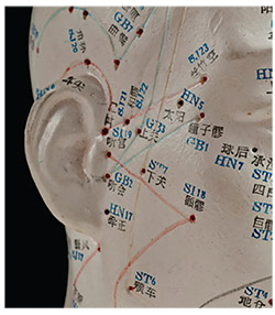 |
| One study found a synergistic effect of acupoint treatment (a model of the various acupoints is shown here) and low-dose atropine in decreasing the progression of myopia. |
• Orthokeratology. Orthokeratology is the use of overnight rigid contact lenses to reshape the cornea. It works by flattening the central cornea, thinning the central corneal epithelium, thickening the mid-peripheral cornea, and producing a myopic shift in the peripheral vision. This temporarily reduces or eliminates refractive error and decreases the need to wear contact lenses or spectacles in the daytime. Researchers found that orthokeratology is as effective as atropine in delaying myopic progression.
Although orthokeratology potentially eliminates the side effects of atropine, it has the potential for significant side effects of its own, including corneal infections, an increase in higher-order corneal aberrations and a decrease in contrast sensitivity.32 Orthokeratology also induces a temporary shift in corneal curvature that returns to baseline after the treatment is stopped. There is limited evidence of slowed long-term axial length changes.
• Refractive undercorrection. Studies in infant monkeys have supported the idea that undercorrection could alter the eye’s growth and therefore change its refractive ability.16 Therapeutic undercorrection may also reduce the near-vision accommodative response which may be a factor in myopic progression. However, when studied, there was no significant difference in myopic progression in children prescribed with undercorrected lenses in comparison to children with fully corrected lenses.17
• Part-time spectacle wear. It’s thought that optical defocus plays an important role in the development of myopia. One study explored the effects of different patterns of lens wear on myopia progression. There was no significant difference in the three-year progression between the full-time lens wearers, those who switched from distance-only to fulltime wear, distance-only wearers and non-wearers.18 This suggests that part-time lens wear is ineffective in the treatment of myopia progression.
• Bifocal and multifocal spectacle correction. It’s been hypothesized that bifocal or multifocal glasses correction could reduce retinal defocus and thus slow myopia progression. However, several clinical trials have shown no significant difference in myopia progression.19,20 One study, though, reported that bifocal-only spectacles and bifocal spectacles with base-in prism slowed myopia progression by 39 percent and 50 percent, respectively, in Chinese-Canadian children.21-23
• Progressive-addition spectacle lenses. Progressive-addition lenses, in comparison to single-vision lenses, were associated with a decrease in myopia progression, but this difference failed to reach clinical significance.24 The authors concluded that although there was a small decrease in myopia progression, this didn’t warrant a change in clinical guidelines.24
• Peripheral retinal defocus. Studies have suggested that the peripheral retina and peripheral vision have roles in the pathogenesis of myopia. In primates, it was found that foveal ablation didn’t have an effect on the emmetropization process.25 The absence of central vision didn’t affect the development of myopia, suggesting that peripheral vision has a more important role. However, in humans it was found that relative peripheral hyperopia had little association with the risk of myopia onset, myopia progression or axial growth.26-29 A study comparing children who wore spectacle lenses that decreased relative peripheral hyperopia to children who wore single-vision spectacle lenses found no significant difference between the two groups. However, in children of myopic parents with higher rates of myopia progression, it was found that correction of relative peripheral hyperopia reduced myopia progression.30
• Pirenzepine. Pirenzepine is a selective M1 muscarinic receptor antagonist. It’s more selective than atropine (discussed below), and therefore results in less cycloplegia and mydriasis.31 It was shown that 12-month use of 2% topical pirenzepine ophthalmic gel twice a day is associated with a 40-percent reduction in axial length.25 Unfortunately, pirenzepine gel isn’t commercially available for use.32 Further studies on pirenzepine’s long-term safety and efficacy are still warranted.
• Atropine. There has been extensive research on the use of atropine in the prevention of myopia progression. Atropine is a nonselective muscarinic antagonist thought to work on the five muscarinic receptor subtypes in the human eye, M1 through M5, and inhibit glycosaminoglycan synthesis in scleral fibroblasts.11,25 Additionally, it’s been proposed that, like light, atropine activates the parasympathetic nervous system through the five muscarinic receptor subtypes found on the human iris sphincter, ciliary body, and throughout the retina, sclera and lens.11 Not only is atropine thought to work directly on the muscarinic receptors in the eye, but it’s also thought to increase or decrease the amount of these receptors.11
Multiple studies in the literature have reported that atropine use has significantly delayed the progression of myopia and axial elongation.6,33-35 Atropine also downregulates inflammatory markers in the eye thought to be involved in myopia progression. Myopic hamsters treated with atropine in the eyes had a decrease in expression of proteins such as c-Fos, NFkB, Il-6 and TNFá, indicating an inflammatory pathway related to myopia.38
Myopic rebound is a concern after discontinuing use of high-dose atropine treatment. However, myopic rebound is seen to a lesser extent with low-dose atropine (0.01%).33 Additionally, slowly tapering the frequency of atropine instead of abruptly stopping treatment might retain the beneficial effect on myopia progression. Immediately stopping high-dose atropine releases its inhibitory effect, causing a growth spurt that contributes to myopia progression.11
Initially, differences in iris color were another source of concern with atropine use. Having a lighter-color iris was considered a contraindication to atropine use due to an increase in reports of adverse events such as photophobia, allergy and poor near vision in such patients.35 However, it was found that similar rates of these adverse events were reported in Asian children with dark-colored irises and Caucasian children with light-colored irises.35 Therefore, light-colored irises should no longer be considered a contraindication to atropine use.
One difficulty with atropine is predicting which children will benefit the most from it. Some evidence has pointed to greater effects of atropine treatment in Asian children in comparison to Caucasians.39 However, atropine has been shown to be effective in many different populations, including Caucasians, Asians and Indians.11,40 More research is needed to better understand the variability in responses to atropine treatment.
It’s also been shown that atropine 0.5%, 0.1% and 0.01% are safe and effective, and there’s no decrease in efficacy with decreasing dosage.35,36 This is important because a lower dosage of atropine is associated with fewer side effects such as poor near vision, loss of accommodation due to cycloplegia and glare, and is also associated with a decreased risk of needing an add power in glasses.36 Additionally, atropine treatment doesn’t have any adverse effects on intraocular pressure, optic nerve parameters or retinal nerve fiber layer thickness.42 Delay in myopic progression and corresponding axial length change by atropine use is thought to be sustained over the long term due to the natural37 slowing of eye growth.
One group of researchers postulated that low-dose atropine produces a sustained response in comparison to high-dose atropine by working more anteriorly in the eye and affecting various muscarinic receptors at different levels.11
There’s been some evidence that combining atropine treatment with another myopia treatment could have an additive effect in decreasing myopia progression. To test this idea, one group of researchers investigated the effects of low-dose atropine treatment with and without auricular acupoint stimulation in myopes.43 They found that patients treated with this combination had less myopic progression, less axial length elongation, more anterior chamber deepening, and greater reductions in intraocular pressure in comparison to patients treated with just atropine.43
Researchers are also studying how to improve atropine delivery to the eye. Two studies have shown that drug-eluting silicone hydrogel soft contact lenses can potentially deliver atropine.44,45 This could eliminate the need for regular drop instillation and improve compliance.
Ongoing studies are looking to answer questions such as which children would benefit most from atropine treatment, the optimal age to begin the treatment, the ideal length of treatment and whether treatments could be combined for better efficacy.
In conclusion, myopia and its associated complications are an increasing public health concern. While glasses and contact lens correction are valuable in treating the symptomatic vision changes associated with myopia, they don’t change the anatomic progression of the myopic eye. Low-dose atropine use (0.01%) remains the most encouraging treatment choice available at this time, though we need more studies to identify its optimal use. Additional environmental studies will also help determine if there are lifestyle changes that could slow myopia’s progression. In the meantime, vision screening and early detection remain essential for diagnosis and correction to avoid the loss of correctable, functional vision. REVIEW
Ms. James is a medical student at the Sidney Kimmel College of Medicine, Thomas Jefferson University in Philadelphia, where Dr. Salvin serves as a clinical associate professor of pediatrics and ophthalmology. Dr. Salvin is also affiliated with the Division of Ophthalmology at the Nemours/ A.I. duPont Hospital for Children in Wilmington, Delaware, and Wills Eye Hospital.
1. Williams KM, Bertelsen G, Cumberland P, et al. Increasing prevalence of myopia in Europe and the impact of education. Ophthalmology 2015;122:7:1489-1497.
2. Vitale S, Sperduto RD, Ferris FL, III. Increased prevalence of myopia in the United States between 1971-1972 and 1999-2004. Archives of Ophthalmology 2009;127:12:1632-1639.
3. Holden BA, Fricke TR, Wilson DA, et al. Global prevalence of myopia and high myopia and temporal trends from 2000 through 2050. Ophthalmology 2016;123:5:1036-1042.
4. Bourne RRA, Stevens GA, White RA, et al. Causes of vision loss worldwide, 1990–2010: A systematic analysis. The Lancet Global Health 2013;1:6:349.
5. Wong TY, Ferreira A, Hughes R, Carter G, Mitchell P. Epidemiology and disease burden of pathologic myopia and myopic choroidal neovascularization: An evidence-based systematic review. American Journal of Ophthalmology 2014;157:1:25.
6. Gong Q, Liu L. Therapeutic effect of atropine 1% in children with low myopia. Journal of American Association for Pediatric Ophthalmology and Strabismus 2016;20:4:379.
7. The Eye Diseases Prevalence Research, Group. The prevalence of refractive errors among adults in the United States, Western Europe and Australia. Archives of Ophthalmology 2004;122:4:495-505.
8. Smith TST, Frick KD, Holden BA, Fricke TR, Naidoo KS. Potential lost productivity resulting from the global burden of uncorrected refractive error. Bulletin of the World Health Organization 2009;87:6:431.
9. Flitcroft DI, Loughman J, Wildsoet CF, et al. Novel myopia genes and pathways identified from syndromic forms of myopia. Investigative Ophthalmology & Visual Science 2018;59:1:338348.
10. Morgan IG, Ohno-Matsui K, Saw S. Myopia. The Lancet 2012;379:9827:1739-1748.
11. Rucker F, Wallman J. Chicks use changes in luminance and chromatic contrast as indicators of the sign of defocus. Journal of Vision 2012;12:6:23.
12. Smith EL, Hung L, Arumugam B. Visual regulation of refractive development: Insights from animal studies. Eye (London, England) 2014;28:2:180.
13. Lim LS, Gazzard G, Low Y, et al. Dietary factors, myopia, and axial dimensions in children. Ophthalmology 2010;117:5:997.
14. Walline JJ, Jones LA, Sinnott L, et al. A randomized trial of the effect of soft contact lenses on myopia progression in children. Investigative Ophthalmology & Visual Science. 2008;49:11:47024706.
15. Walline JJ, Jones LA, Mutti DO, Zadnik K. A randomized trial of the effects of rigid contact lenses on myopia progression. Archives of Ophthalmology 2004;122:12:1760-1766.
16. Smith III EL, Hung L. The role of optical defocus in regulating refractive development in infant monkeys. Vision Research 1999;39:8:1415-1435.
17. Adler D, Millodot M. The possible effect of undercorrection on myopic progression in children. Clinical & Experimental Optometry 2006;89:5:315-321.
18. Ong E, Grice K, Held R, Thorn F, Gwiazda J. Effects of spectacle intervention on the progression of myopia in children. Optometry and Vision Science 1999;76:6:363-369.
19. Jensen H. Myopia progression in young school children. A prospective study of myopia progression and the effect of a trial with bifocal lenses and beta blocker eye drops. Acta ophthalmologica (Supplement) 1991:200:1.
20. Pärssinen O, Hemminki E, Klemetti A. Effect of spectacle use and accommodation on myopic progression: Final results of a three-year randomised clinical trial among schoolchildren. The British Journal of Ophthalmology 1989;73:7:547-551.
21. Cheng D, Woo GC, Drobe B, Schmid KL. Effect of bifocal and prismatic bifocal spectacles on myopia progression in children: Three-year results of a randomized clinical trial. JAMA Ophthalmology 2014;132:3:258-264.
22. Cheng D, Schmid KL, Woo GC, Drobe B. Randomized trial of effect of bifocal and prismatic bifocal spectacles on myopic progression: Two-year results. Archives of Ophthalmology 2010;128:1:12-19.
23. Cheng D, Woo GC, Schmid KL. Bifocal lens control of myopic progression in children. Clinical & Experimental Optometry 2011;94:1:24.
24. Gwiazda J, Hyman L, Hussein M, et al. A randomized clinical trial of progressive addition lenses versus single vision lenses on the progression of myopia in children. Investigative Ophthalmology & Visual Science. 2003;44:4:1492.
25. Leo SW, Scientific Bureau of World Society of Paediatric Ophthalmology and Strabismus (WSPOS). Current approaches to myopia control. Curr Opin Ophthalmol 2017;28:3:267-275.
26. Smith EL, Ramamirtham R, Qiao-Grider Y, et al. Effects of foveal ablation on emmetropization and form-deprivation myopia. Investigative Ophthalmology & Visual Science 2007;48:9:3914.
27. Mutti DO, Hayes JR, Mitchell GL, et al. Refractive error, axial length, and relative peripheral refractive error before and after the onset of myopia. Investigative Ophthalmology & Visual Science 2007;48:6:2510.
28. Mutti DO, Sinnott LT, Mitchell GL, et al. Relative peripheral refractive error and the risk of onset and progression of myopia in children. Investigative ophthalmology & visual science 2011;52:1:199.
29. Sng CCA, Lin X, Gazzard G, et al. Change in peripheral refraction over time in Singapore Chinese children. Investigative Ophthalmology & Visual Science 2011;52:11:7880.
30. Sankaridurg P, Donovan L, Varnas S, et al. Spectacle lenses designed to reduce progression of myopia: 12-month results. Optometry and Vision Science 2010;87:9:631-641.
31. Huang J, Wen D, Wang Q, et al. Efficacy comparison of 16 interventions for myopia control in children: A network metaanalysis. Ophthalmology 2016;123:4:697-708.
32. Galvis V, Tello A, Parra MM, et al. Five-year clinical trial on atropine for the treatment of myopia 2: Myopia control with atropine 0.01% eyedrops. Ophthalmology 2016;123:6:41.
33. Chia A, Chua W, Wen L, Fong A, Goon YY, Tan D. Atropine for the treatment of childhood myopia: Changes after stopping atropine 0.01%, 0.1% and 0.5%. American Journal of Ophthalmology 2014;157:2:457.
34. Pineles SL, Kraker RT, VanderVeen DK, et al. Atropine for the prevention of myopia progression in children: A report by the American Academy of Ophthalmology. Ophthalmology 2017;124:12:1857-1866.
35. Gong Q, Janowski M, Luo M, et a. Efficacy and adverse effects of atropine in childhood myopia: A meta-analysis. JAMA Ophthalmology 2017;135:6:624-630.
36. Chia A, Chua W, Cheung Y, et al. Atropine for the treatment of childhood myopia: Safety and efficacy of 0.5%, 0.1%, and 0.01% doses (atropine for the treatment of myopia 2). Ophthalmology 2012;119:2:347-354.
37. Chia A, Lu Q, Tan D. Five-year clinical trial on atropine for the treatment of myopia 2: Myopia control with atropine 0.01% eyedrops. Ophthalmology 2016;123:2:391-399.
38. Lin H, Wei C, Chang C, et al. Role of chronic inflammation in myopia progression: Clinical evidence and experimental validation. EBioMedicine 2016;10:269-281.
39. Li S, Wu S, Kang M, et al. Atropine slows myopia progression more in Asian than white children by meta-analysis. Optometry and Vision Science 2014;91:3:342-350.
40. Kothari M, Rathod V. Efficacy of 1% atropine eye drops in retarding progressive axial myopia in Indian eyes. Indian Journal of Ophthalmology 2017;65:11:1178-1181.
41. Chan L, Hsieh Y, Hsu W, Cheng H, Shen EP. Optic disc parameters of myopic children with atropine treatment. Curr Eye Res 2017;42:12:1614-1619.
42. Stone RA, Pardue MT, Iuvone PM, Khurana TS. Pharmacology of myopia and potential role for intrinsic retinal circadian rhythms. Experimental Eye Research 2013;114:35-47.
43. Cheng H, Hsieh Y. The effect of low-concentration atropine combined with auricular acupoint stimulation in myopia control. Complementary Therapies in Medicine 2014;22:3:449-455.
44. Hui A, Bajgrowicz-Cieslak M, Phan CM, Jones L. In vitro release of two anti-muscarinic drugs from soft contact lenses. Clinical Ophthalmology 2017:1657-1665.
45. Lasowski F, Sheardown H. Atropine and roscovitine release from model silicone hydrogels. Optometry and Vision Science 2016;93:4:404-411.


