A simple eye test may someday offer an effective way to identify patients who are at high risk for stroke, say researchers at the University of Zurich. They showed that a test called ocular pulse amplitude (OPA) can reliably detect carotid artery stenosis (CAS), a known risk factor for stroke. The OPA test could be performed by ophthalmologists during routine exams. The study, published in the
June Ophthalmology, confirmed that patients who had the lowest OPA scores also had the most seriously blocked arteries.
Each year, approximately 795,000 Americans suffer a new or recurrent stroke, and more than 137,000 of these people die as a result. People with severe CAS are much more likely to suffer stroke. Physicians would like to catch and treat CAS before that can happen, but because CAS has no symptoms and an efficient test is not currently available, the disease often goes undetected.
The Swiss research team used a device called the dynamic contour tonometer to check the OPA of 67 patients who were assumed to have CAS. The OPA score is calculated by finding the difference between the intraocular pressure levels during the systolic and diastolic phases of the heartbeat. The tonometer measures the two pressure levels, then instantly computes the patient’s OPA score. When blood flow to the eye is blocked by CAS, there is not much difference between the two pressure levels, so the OPA score is low. The study confirmed that patients with the lowest OPA scores also had the most seriously blocked arteries. The researchers used ultrasound exams to corroborate that each study participant had CAS and to detail the severity of the blockage.
“Our results show that ocular pulse amplitude is a reliable, safe screening test for carotid artery stenosis,” said lead researcher Pascal Bruno Knecht, MD. “We recommend further study to confirm the value of using OPA to detect and assess the severity of CAS and to define its use in stroke prevention.”
A research review performed for the U.S Preventive Services Task Force indicated that if an efficient screening test for CAS were available, the incidence of stroke and fatalities due to stroke could be substantially reduced. The review stated that the test should be able to detect clinically significant CAS, defined as 60 percent to 99 percent blockage of the carotid arteries. Some high-tech tests, such as magnetic resonance angiography and color duplex ultrasound, already meet this standard, but they are expensive and not widely available. Their primary use is in diagnosing patients who already have symptoms of stroke.
It could be efficient to perform the OPA test during a standard eye exam, if the ophthalmologist is already using the dynamic contour tonometer to screen for glaucoma. This type of tonometer is not widely used in the United States, although it is in Europe.
The researchers say that other than CAS, very few diseases could cause low OPA scores, and that an ophthalmologist could easily rule out these other diseases during an eye exam.
Lenses May Slow Myopia Progress in Children
Research at the University of Houston College of Optometry suggests that optical treatments warrant further study for their potential to slow the progression of nearsightedness in children.
Conducted by UH assistant professor David Berntsen, OD, PhD, and his colleagues from Ohio State University, the study compared the effects of wearing and then not wearing progressive addition lenses in children who are nearsighted. The study examined 85 children from 6 to 11 years old over the course of two years. The results were published in
Investigative Ophthalmology and Visual Science.
Selected according to their eye alignment and accuracy of focusing on near objects, the myopic children were fitted with either normal single-vision lenses or no-line bifocals to correct their nearsightedness. In addition to observing and testing the children, the doctors obtained feedback from parents and guardians of both the children’s outdoor activities and near-work tasks, such as reading and computer use.
Previous research suggested that nearsighted children who do not focus accurately when reading books or doing other near work may benefit more from wearing no-line bifocal glasses than nearsighted children who focus more accurately. Dr. Berntsen’s study found a small, yet statistically significant, slowing of myopia progression in children wearing the bifocals compared to those who simply wore single-vision lenses. Dr. Berntsen asserts, however, that the results do not suggest that children be fitted with no-line bifocal lenses solely for the purpose of slowing the progression of myopia.
“While the small effect found in the group of children wearing bifocal spectacles does not warrant a change in clinical practice, we found the beneficial effect was still present for at least one year after children stopped wearing no-line bifocal lenses,” Dr. Berntsen said. “This is promising if other optical lens designs can be developed that do an even better job of slowing how fast myopia increases in children.”
By understanding why different types of lenses result in the slowing of myopia progression, Dr. Berntsen says researchers will be better able to design lenses that may be more effective in slowing the increase of nearsightedness in children.
“Single-vision lenses are normally prescribed when a child gets a pair of glasses, but glasses with progressive addition lenses were shown to slightly reduce myopic progression in our study,” he said. “For any treatment that reduces myopia progression in children to be useful, the effect of the spectacles or contact lenses must persist after children stop wearing them. The fact that the small treatment effect from our study was still present one year after discontinuing the treatment is promising. The results suggest that if newer optical designs currently being investigated do a better job of slowing myopia progression, the effects may be expected to persist and decrease how nearsighted the child ultimately becomes.”
Dr. Berntsen says the study results and evidence from other studies suggest that lenses specifically designed to change blur in the eye’s peripheral vision may be able to slow the increase of nearsightedness.
“There is support for continuing to investigate new lenses specially designed to change the blur profile on the back of the eye in order to reduce the increase of myopia in children,” he said. “There is still further research to be done, but our work is an important step in discovering the methods needed to slow the progression of nearsightedness.”
|
Stem Cells Able to Form Optic Cup
Human-derived stem cells can spontaneously form an optic cup, according to a study published in
Cell Stem Cell. Transplantation of this 3D tissue in the future could help patients with visual impairments see clearly.
“This is an important milestone for a new generation of regenerative medicine,” says senior study author Yoshiki Sasai, MD, PhD, of the RIKEN Center for Developmental Biology, in Kobe, Japan. “Our approach opens a new avenue to the use of human stem cell-derived complex tissues for therapy, as well as for other medical studies related to pathogenesis and drug discovery.”
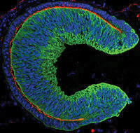
In the study, the optic cup spontaneously emerged from human embryonic stem cells (hESCs)—cells derived from human embryos that are capable of developing into a variety of tissues—thanks to the cell culture methods optimized by Dr. Sasai and his team.
The hESC-derived cells formed the correct 3D shape and the two layers of the optic cup, including a layer containing a large number of light-responsive photoreceptor cells. Because retinal degeneration primarily results from damage to these cells, the hESC-derived tissue could be ideal transplantation material.
Beyond the clinical implications, the study will likely accelerate the acquisition of knowledge in the field of developmental biology. For instance, the hESC-derived optic cup is much larger than the optic cup that Dr. Sasai and collaborators previously derived from mouse embryonic stem cells, suggesting that these cells contain innate species-specific instructions for building this eye structure. “This study opens the door to understanding human-specific aspects of eye development that researchers were not able to investigate before,” Dr. Sasai says.
HSV Infection Tied to AMD
A team of researchers reports that human cytomegalovirus, a type of herpes virus, is associated with neovascular age-related macular degeneration. They report that human cytomegalovirus causes the production of vascular endothelial growth factor, a signal protein that regulates the formation of new blood vessels. The results were published in
PLoS Pathogens.
“Prior to this work, cofactors for the development of AMD included genetics, a high-fat diet and smoking. Now, we are adding an infectious agent as another cofactor,” said Richard D. Dix, professor at the Georgia State Viral Immunology Center’s Ocular Virology and Immunology Laboratory. Affiliated research institutions include the Duke University Eye Center, the Bascom Palmer Eye Institute of the University of Miami Miller School of Medicine, the Viral Immunology Center at Georgia State, and the Department of Ophthalmology at the Emory University School of Medicine.
Human cytomegalovirus is a common herpes virus, said Dr. Dix. About 80 percent of the population is estimated to have antibodies for the virus, and it is often acquired during childhood. In a normal, healthy immune system, the virus becomes latent in the cells of bone marrow and blood. But in the elderly, the immune system’s function is reduced, the virus proliferates and the production of VEGF increases.
Identifying human cytomegalovirus as a cofactor in the development of AMD opens up new paths for the treatment of AMD, Dr. Dix said. One route could include reducing the viral load—the amount of the human cytomegalovirus in the blood stream—by treatment with an antiviral drug known as ganciclovir.
Additional research paths include looking at the genetics involved in the upregulation of VEGF by human cytomegalovirus. “If we can knock down a certain gene or genes of the virus that stimulates VEGF production, we might be able to decrease its production and minimize AMD,” Dr. Dix said. REVIEW


