In a total of 118 consecutive eyes of 118 patients who underwent uneventful DMEK for Fuchs’ endothelial dystrophy at a tertiary referral center, best spectacle-corrected visual acuity, corneal HOAs and backscattered light were evaluated preoperatively and at six months postop. Outcome data was compared to an age-matched control group with uncomplicated eyes (n=27).
Compared to the control group, Fuchs’ eyes, before as well as six months after DMEK, showed higher values of anterior and posterior HOAs and backscattered light (p<0.033). Postoperative anterior HOAs and backscattered light (0 to 2 mm) were associated with lower six-month BCSVA (positively related with logMAR BSCVA; p≤0.02). Anterior corneal HOAs did not change from preop to six months after DMEK (p=0.649), while total posterior HOAs (RMS third to sixth Zernike order) and haze decreased (p<0.001). If present, anterior surface irregularities and anterior corneal haze may be the most important limiting factors in visual rehabilitation after DMEK for eyes suffering from Fuchs’ endothelial dystrophy.
Am J Ophthalmol 2014;158:71-79.
Van Dijk K, Droutsas K, Hou J, Sangsari S, et al.
Culture and Adherence to Glaucoma Treatment
In an effort to determine adherence rates and beliefs about glaucoma and its treatments, researchers examined 475 patients using topical eye drops for at least six months in a cross-sectional study. The sample consisted of white Americans (n=133), African Americans (n=58), white Australians (n=107) and Singaporeans of Chinese descent (n=117).
 |
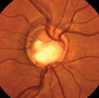
Accounting for sociodemographic differences, significant differences in self-reported adherence rates were identified (p<0.001). White Americans (65.4 percent) and Australians (67.7 percent) reported significantly higher adherence than African Americans (56.9 percent) or Singaporeans (47.5 percent). Beliefs about glaucoma treatment were predictive of adherence only in Australian and white American samples (p<0.06). This suggests that in Western cultures, attempts to improve adherence may benefit from greater examination of individuals’ concerns about, and perceived need of, glaucoma treatment.
J Glaucoma 2014;23:293-298.
Rees W, Chong X, Cheung C, Aung T, Friedman D, et al.
Two-year Results from the COPERNICUS Study
New research from the COPERNICUS study, which evaluated the efficacy and safety of intravitreal aflibercept injection for the treatment of macular edema secondary to central retinal vein occlusion, shows that the visual and anatomic improvements after fixed dosing through week 24 and p.r.n. dosing with monthly monitoring from weeks 24 to 52 were diminished after continued p.r.n. dosing with a reduced monitoring frequency from weeks 52 to 100.
A total of 188 patients with macular edema secondary to CRVO were placed in a randomized, double-masked Phase III trial. Patients (n=114) received IAI 2 mg (IAI 2Q4) or sham injections (n=74) every four weeks up to week 24. During weeks 24 to 52, patients from both arms were evaluated monthly and received IAI as needed (IAI 2Q4 + p.r.n. and sham + IAI p.r.n.). During weeks 52 to 100, patients were evaluated at least quarterly and received IAI as needed.
The proportion of patients gaining ≥15 letters of best-corrected visual acuity was 56.1 percent vs. 12.3 percent (p<0.001) at week 24, (the primary COPERNICUS study efficacy endpoint); 55.3 percent vs. 30.1 percent (p<0.001) at week 52; and 49.1 percent vs. 23.3 percent (p<0.001) at week 100 in the IAI 2Q4 + p.r.n. and sham + IAI p.r.n. groups, respectively. The mean change from baseline BCVA was also significantly higher in the IAI 2Q4 + p.r.n. groups compared with the sham + IAI p.r.n. group at week 24 (+17.3 vs. -4.0 letters; p<0.001); week 52 (+16.2 vs. 3.8 letters; p<0.001); and week 100 (+13.0 vs. 1.5 letters; p<0.001). The mean reduction from baseline in central retinal thickness was 457.2 vs. 144.8 μm (p<0.001) at week 24, 413 vs. 381.8 μm at week 52 (p=0.546) and 390 vs. 343.3 μm at week 100 (p=0.366) in the IAI 2Q4 + p.r.n. and sham + IAI p.r.n. groups. The mean number of p.r.n. injections in the IAI 2Q4 + p.r.n. and sham + IAI p.r.n. groups was 2.7 ±1.7 vs. 3.9 ±2.0 during weeks 24 to 52 and 3.3 ±2.1 vs. 2.9 ±2.0 during weeks 50 to 100. The most frequent ocular serious adverse event from baseline to week 100 was vitreous hemorrhage (0.9 percent vs. 6.8 percent in the IAI 2Q4 + p.r.n. and sham + IAI p.r.n. groups).
Ophthalmology 2014;121:1414-1420.
Heier J, Clark WL, Boyer S, Brown D, Vitti R, et al.
One-year Follow-up of Accelerated CXL for Keratoconus
Researchers from the University of Toronto assessed the efficacy of accelerated corneal collagen cross-linking (irradiance of 9mW/cm2; 10 minutes) in keratoconus-affected eyes, determining that it is effective in stabilizing topographic parameters after 12 months of follow-up. Improvement in the uncorrected distance visual acuity and stabilization of all tested corneal parameters were noted after the treatment.
Sixteen mild-moderate keratoconus-affected eyes of 14 patients (mean age: 24.9 ±5.8 years; range: 17.1 to 38.3 years) who underwent accelerated CXL treatment and had six and 12 months of follow-up were reviewed retrospectively. Data regarding UDVA, manifest refraction, corrected distant visual acuity and computerized corneal topography data before and post-CXL surgery were extracted and analyzed.
No statistically significant changes were found in the mean CDVA, mean refractive cylinder or mean manifest refraction spherical equivalent at either time point. There was a gain of 0.13 logMAR in the mean UDVA (p=0.012) at 12 months. All corneal parameters including Ksteep, Kflat, average K (Km), corneal astigmatism (Kcy1) and maximal curvature reading the corneal apex (Kmax) were stable at six and 12 months in all patients. No complications were observed during the follow-up period.
Cornea 2014;33:769-773.
Elbaz U, Shen C, Lichtinger A, Zauberman N, et al.


