Gene Therapy Pushes Forward
One of the most exciting areas of current ophthalmic research is gene therapy. Scientists and clinicians are taking advantage of the unique features of the visual system to make the first efforts at correcting genetic ophthalmic disorders. A prime target for these efforts is choroideremia, a single-locus, x-linked condition that results in a progressive loss of vision over several decades; affected individuals have significant loss of night vision by the second decade and are legally blind by 40 to 50 years of age.1,2 The disease is due to the absence of the REP1 gene product, a choroid protein involved in post-translational prenylation. The slow rate of visual decay and small size of the affected gene make this condition an ideal test case for gene therapy. A research group headed by Dr. Robert MacLaren (Oxford Eye Hospital; Oxford, U.K.) recently published results from six patients treated with a functional copy of the REP1 gene packaged in an adeno-associated viral vector designed for choroidal expression. All six patients showed significant increases in VA.3
At ARVO, experimental details and obstacles to effective therapy were discussed in a series of presentations. (Fischer M, et al. ARVO E-Abstract 6001; MacLaren R, et al. ARVO E-Abstract 832) A study aimed at screening for immune responses to the vector or the rescue gene product was also presented, (Barnard A, et al. ARVO E-Abstract 3296) while another presentation focused on the two subjects out of six from Dr. MacLaren’s study who had more advanced impairment, including retinal detachment. (Groppe M, et al. ARVO E-Abstract 3295) Despite these potential limitations, both subjects tolerated the viral injections without adverse effects and showed significant increases in visual acuity.
|
Stepping back from the clinic for a more long-term view, we were excited by a number of presentations on progress that’s being made with induced pluripotent stem cells. The logic behind this approach, at least in part, is that a patient’s own cells are the best targets for reprogramming with a corrected gene. These cells are then induced to develop into the tissue of choice. Introduction of the genetic correction involves one of several DNA editing schemes followed by transplantation of repaired cells.5 Examples of this approach were described for a therapy to treat Knoblach’s syndrome (Nguyen H, et al. ARVO E-Abstract 2982), as well as a test treatment to correct the male germ cell-associated kinase mutation that causes retinitis pigmentosa in patients of Ashkenazi Jewish ancestry. (Stone E, et al. ARVO E-Abstract 2676) Edwin Stone, MD, of the University of Iowa, showed that a corrected MAK was functionally expressed in iPSCs, a key step in bringing this technology to the clinic.
Swept-Source OCT
ARVO is a particularly opportune time to explore the latest in imaging technology. This year the biggest buzz seemed to focus on comparisons between spectral-domain and swept-source optical coherence tomography.6 As these technologies evolve, there seems to be a progression of applications as well, as OCT use for imaging anterior structures expands as it has for the retina.
Several reports provided comparisons of retinal nerve fiber layer thickness measurement using SD and SS OCT. (Ha A, et al. ARVO E-Abstract 4741; Lee B, et al. ARVO E-Abstract 3347) In most of these studies, comparison of measures from SS and SD showed significant differences, suggesting values from the two methods are not readily comparable without further standardization. Another study compared retinal imaging by SD and SS in control eyes and in eyes with various opacities, including those with cataracts, vitreous opacity or corneal opacity. (Shin Y, et al. ARVO E-Abstract 3359) The captured images were subjectively graded by two retina specialists using a standardized OCT grading system. While images from normal eyes obtained by either method weren’t significantly different, SS provided a significant improvement over SD in all eyes with reduced opacity.
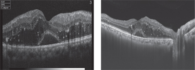
Another area of considerable interest was highlighted by studies that added a time component to SS-OCT to generate 4-dimensional images. One such study (Migacz J, et al. ARVO E-Abstract 5019) employed a technique termed phase variance OCT to image chorioretinal vascular flow. This study showed that the technique may provide greater depth resolution than traditional fluorescein angiography. Other presentations that added a time component to SS-OCT were aimed at integrating this imaging modality into the operating theater to track surgical maneuvers in real time. (Carrasco-Zevallos O, et al. ARVO E-Abstract 1633; Keller B, et al. ARVO E-Abstract 1631) These studies suggest that such high-resolution, high-speed imaging devices may be part of the operating rooms of the future.
Two reports used SS-OCT in assessments of angle dimensions and lamina cribrosa insertion in open-angle glaucoma. (Rigi M, et al. ARVO E-Abstract 930; Lee K, et al. ARVO E-Abstract 904) The resolution of the swept-source devices allows for precise morphometric analysis and provides support for the growing emphasis of these imaging metrics in the diagnosis of glaucoma. In a related study, defects in the lamina cribrosa were compared in myopes with or without glaucoma along with normal controls. (Miki A, et al. ARVO E-Abstract 908) This pilot study confirmed reports that such defects may be associated with development of the disease. The number of eyes with at least one focal defect in the LC were significantly different between groups: 1/20 in normal eyes; 6/32 in myopes; and 27/66 in the eyes with both myopia and glaucoma.
SS-OCT is also being employed for anterior-segment imaging, as described in a study comparing corneal thickness measurements determined by ultrasonic pachymetry and anterior segment tomography with those obtained by SS-OCT. (Haines L, et al. ARVO E-Abstract 2464) Although the sample size was small (n=16 eyes), the study showed all devices provided comparable measures. The authors point out that “in addition to high resolution morphological imaging of the cornea, SS-OCT can provide precise morphometric analysis of the human cornea.”
Several head-to-head comparisons of devices from different manufacturers also provided a key perspective. Heidelberg Spectralis SD-OCT and Topcon Deep Range Imaging SS-OCT for macular imaging were used on the same subjects, providing a concise depiction of relative strengths and limitations of each device. (Barteselli G, et al. ARVO E-Abstract 360) Authors summarized their results by concluding that while “details of the pre-retinal vitreous are better imaged using the shorter wavelength of the spectral domain OCT, the sharpness of the choroidal structures is better using the higher scanning speed of the SS-OCT.” They also point out that the two devices are comparable for visualizing the choroidal border, and that these are the images that are used to generate choroidal thickness measurements.
OCT has been used in published studies for measurement of tear meniscus height, but the higher speed and resolution of SS-OCT allow for unique studies of meniscus dynamics. (Fukuda IOVS 2014 55: ARVO E-abstract 1981) In a trial of 23 subjects, all with normal values for tear-film breakup, Schirmer’s test and corneal fluorescein staining, a time course of tear meniscus height and volume was generated following instillation of saline, sodium hyaluronate or rebamipide. Time points ranged from 30 seconds to 20 minutes after instillation. Significant increases in meniscus measures were observed until one minute post-instillation for saline, three minutes post-instillation for 0.1% sodium hyaluronate, 10 minutes post-instillation for 0.3% sodium hyaluronate, and five minutes post-instillation for rebamipide. The authors suggest that SS-OCT could provide a new metric in studies of eye-drop efficacy.
Tear dynamics are always a topic of interest. One study examined expression of Muc16, an important mucin component of the tear film. Muc16 is expressed by conjunctival and corneal apical epithelium cells, which shed the extracellular domain of the protein into the surrounding tear layer. In a methodical series of experiments, Ilene Gipson and colleagues showed that goblet cells also produce a variant of Muc16 and contribute this to the tear film in both mouse and human eyes. (Gipson IOVS 2014 55: ARVO E-Abstract 2760) This finding is noteworthy for a number of reasons, but particularly because it suggests a greater than previously appreciated role for goblet cells in tear-film homeostasis.
Therapeutic Highlights
For us, the bread and butter of ARVO are the presentations on new therapeutics, both pre-clinical and clinical. Among the preclinical studies was a description of a new type of antihistamine with mixed receptor specificity. (Chapin M, et al. ARVO E-Abstract 2482) These new compounds, GD135 and GD136 (Griffin Discoveries; Amsterdam, Netherlands), antagonize both H1 histamine receptors (the target of traditional antihistamines) and the H4 receptor, which is thought to be important in various signal-processing pathways, including those involving pruritis.7
|
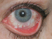
Several presentations described clinical studies of topical antihistamines for allergic conjunctivitis. Aciex Therapeutics presented a study of its topical formulation of cetirizine, AC-170, an antihistamine commonly used in oral formulations but not yet available for topical use. The formulation was shown to significantly and rapidly reduce ocular itch, lid swelling and other signs of ocular allergy. (Gomes P, et al. ARVO E-Abstract 2490) Alcon presented Phase III results for olopatadine 0.77%, a new higher-strength formulation of the topical antihistamine currently available as Pataday or Patanol, that confirmed that this new formulation is superior to placebo at both 16 and 24 hours, providing unqualified q.d. dosing for ocular itching. (McLaurin E, et al. ARVO E-Abstract 2488)
The anti-proliferative agent PRI-321 (Prism Pharma; King of Prussia, Pa.) holds promise as an anti-fibrotic treatment for conditions such as choroidal neovascularization or proliferative vitreoretinopathy. (Whitlock A, et al. ARVO E-Abstract 1203) In a laser-induced model of CNV, Prism Pharma researchers and others found PRI-321 to be as good as an anti-VEGF comparator. A retinal detachment model of PVR showed similar results, with PRI-321 significantly reducing Müller cell proliferation and scar formation. Data on another new compound of interest came from Amakem’s (Diepenbeek, Belgium) successful first-in-human Phase I/Phase II trial of AMA0076, a Rho kinase inhibitor with properties that minimize adverse effects without impacting efficacy. (Hall J, et al. ARVO E-Abstract 565) Another report of a successful Phase I/Phase II trial came from Aerpio, whose drug AKB-9778 is showing promise as an alternative to VEGF inhibitors for DME. (Brigell M, et al. ARVO E-Abstract 1757)
In terms of new therapies, biomarkers and diagnostic approaches, there is never a shortage of presentations on dry eye. A series of posters describe our work at Ora in refining our ability to quantify and characterize blink behavior and pathophysiology. One study characterized our continuous, automated blink monitoring device, confirming that it provides valid metrics of blink dynamics. (Rodriguez J, et al. ARVO E-Abstract 3681) A second study then used the device to reveal dramatic differences in blinking behavior in subjects who wore either spectacle or contact lens correction. (Heckley C, et al. ARVO E-Abstract 6062) This study also showed large differences in blink between lens products, suggesting that blink monitoring may be a useful metric in lens development.
A number of studies presented approaches to ocular surface pathology, exploring novel methods to assess the corneal and conjunctival insults that occur in chronic allergy, dry eye and other conditions. One study correlated tear fluid biomarkers with various subgroups of dry-eye patients, and uncovered a strong correlation between the tear protein PRR4 and aqueous-deficient dry eye. (Perumal N, et al. ARVO E-Abstract 2002) Another group examined the utility of matrix-metalloproteinase-9 assays in tear samples from dry-eye patients. (Messmer E, et al. ARVO E-Abstract 2001) Objective assessments of dry eye are notorious for their lack of correlation, but this group found that MMP-9 levels in 101 subjects showed a strong positive correlation with OSDI scores, tear-film breakup times, Schirmer’s scores and ocular surface staining. In addition, levels were significantly increased in females, subjects with autoimmune or thyroid disease and those who identified themselves as having Sjögren’s syndrome. Combined with the simplicity of the assay, these findings suggest that MMP-9 may be a valid metric for both clinical diagnosis and drug development applications.
An animal model using an injection of concanavalin A into the lacrimal gland may improve upon the conventional scopolamine-based model of dry eye. (Belen L, et al. ARVO E-Abstract 3663) Transiently elicited decreases in tearing and increases in corneal staining were reduced by oral dexamethasone, suggesting that dry-eye symptomology generated by ConA injection is modifiable and thus useful in testing novel dry-eye therapies.
One study found an increased incidence in signs of dry eye in patients with diabetic peripheral neuropathy when compared to age-matched controls. (DeMill D, et al. ARVO E-Abstract 1483) While the investigators saw no clear association between the severity of the two diseases, dry-eye signs (osmolarity, Schirmer’s test) were significantly higher in diabetic neuropathy patients. As with other types of dry eye, there was a lack of association between signs and OSDI-based symptoms.8
A number of studies examined factors thought to contribute to dry-eye disease. One group examined tear cytokines before and after computer use. (Kumar N, et al. ARVO E-Abstract 1859) Even after only an hour of exposure, increases in galectin-3 and epithelial expression of MMP9 indicated the presence of surface inflammation. Though the study was small (n=5), results suggest that the inflammatory effects of ocular stress occur within a very short time period. Another study compared 20 patients with dry eye to 20 age-matched controls using a driving simulator in combination with traditional DE metrics. (Deschamps N, et al. ARVO E-Abstract 1987) Subjects with DE show significantly longer reaction times and a decreased ability to avoid randomly displayed targets.
One of the biggest issues with dry-eye patients is reading, which is a major contributor to visual stress and ocular discomfort, while at the same time being a key aspect of a patient’s quality of life. It’s not surprising that impairment of reading is one of most important reasons patients seek help for their dry eye. Several studies examined this issue to understand how dry eye impacts reading function, assess these effects and determine the extent to which reading function can be used as a metric in dry-eye studies. A pilot study compared results of reading tests such as the Wilkins test and the IreST test in normals and in dry-eye patients and found a clear pattern of reduced scores in those with dry eye. (Ousler G, et al. ARVO E-Abstract 160) Another study surveyed patients to assess how dry eye impacted their reading tasks. (Watson M, et al. ARVO E-Abstract 1997) As a critical issue of quality of life, it seems that reading might be an appropriate clinical metric for dry-eye therapies in the future.
We enjoyed our visit to Orlando, and hope that next year’s meeting can match the variety of science, technology and therapeutics at ARVO 2014. If past experience is any predictor of what’s ahead, ARVO 2015 in Denver will not disappoint. REVIEW
Dr. Abelson is a clinical professor of ophthalmology at Harvard Medical School.
1. Kalatzis V, Hamel CP, MacDonald IM. First International Choroideremia Research Symposium. Choroideremia: Towards a therapy. Am J Ophthalmol 2013;156:433-7.
2. Huckfeldt RM, Bennett J. Promising first steps in gene therapy for choroideremia. Hum Gene Ther 2014;25:2:96-7.
3. MacLaren RE, Groppe M, Barnard AR, et al. Retinal gene therapy in patients with choroideremia: Initial findings from a phase I/II clinical trial. Lancet 2014;383:9923:1129-37.
4. Dalkara D, Sahel JA. Gene therapy for inherited retinal degenerations. C R Biol 2014;337:185-92.
5. Tucker BA, Mullins RF, Stone EM. Stem cells for investigation and treatment of inherited retinal disease. Human Mol Gen 2014;R1–R8.
6. Adhi M, Duker JS. Optical coherence tomography—current and future applications. Curr Opin Ophthalmol 2013;24:3:213-21.
7. Thurmond RL, Gelfand EW, Dunford PJ. The role of histamine H1 and H4 receptors in allergic inflammation: The search for new antihistamines. Nat Rev Drug Discov 2008;7:41-53.
8. Walker PM, Lane KJ, Ousler GW 3rd, Abelson MB. Diurnal variation of visual function and the signs and symptoms of dry eye. Cornea 2010;29:6:607-12.


