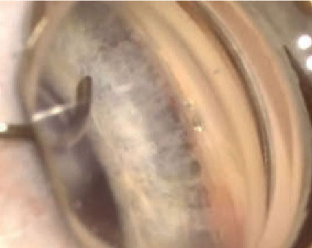
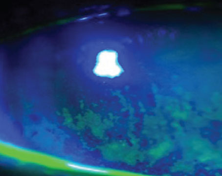
The role of central corneal thickness in glaucoma management is not completely understood, but several interesting aspects have come to light. This article reviews key developments in our understanding on the role of CCT in glaucoma diagnosis and management.
Growing Evidence
In the 1970s, Niels Ehlers, MD, and colleagues reported that patients with low-tension glaucoma had thin corneas1 and individuals with ocular hypertension had thick corneas.2 Over time, other factors were associated with thin CCT: black race,3 old age,4 myopic refractive errors4 and diabetes.4
Most recently, the Ocular Hypertension Treatment Study found that subjects with CCT of <555 µm had a three-fold greater risk for developing primary open angle glaucomae5 Patients with OHT and thin corneas may also demonstrate early visual field defects when tested with short wave automated perimetry.6 Similarly, patients with thin corneas may respond to topical therapy differently, as such individuals show greater measured response to therapy.7
As shown in Table 1, several studies have demonstrated a wide range of CCT measurements in normal subjects and patients with primary open-angle glaucoma, OHT or normal- tension glaucoma. This has put the standard measurement of intraocular pressure by Goldmann applanation tonometry (GAT) into question because GAT assumes a constant CCT of 520 µm.15 Therefore, in an individual with a thick cornea, IOP measurement by GAT may show falsely high reading and the opposite may be true if the cornea is thinner than 520 µm. In a manometric study, Dr. Ehlers calculated GAT to be accurate at 520 µm, but a variation of 70 µm in thickness or thinness resulted in IOP change by 5 mmHg correspondingly.2
Similarly, a meta analysis review on CCT studies16 determined that every 25 µm of change in CCT from a mean of 530 µm resulted in overestimation or underestimation of IOP by 1 mmHg. Another indication of the importance of CCT on IOP measurement is the ever-increasing popularity of corneal refractive surgery. Both photorefractive keratectomy and LASIK result in lower IOP readings after surgery.17,18 IOP measurement by pneumotonometry (PT) on the other hand is less adversely affected by corneal thickness or curvature, which may explain its relative accuracy after keratorefractive surgery.19 Several investigators have observed that PT readings tend to be 1 to 2 mmHg higher than GAT.20,21
Challenging Goldmann
In 2001, a study was undertaken at our institution to determine the effect of race and gender on CCT in patients with POAG and OHT. IOP was measured by GAT and PT. Exclusion criteria were: refractive error of >3D, astigmatism of >1D, prior intraocular procedure, corneal abnormality, and secondary glaucoma. The study enrolled 278 patients, and the results are tabulated in Table 2 and Figure 1. CCT was affected by race: Caucasians had thick corneas, African-Americans had thin corneas and Hispanics had values in between the two.
| Table 1. The Chief Studies on Central Corneal Thickness | ||||||
| Author/Year | Country | Race | CCT (um) | |||
| Normal | POAG | NTG | OHT | |||
| Barbados Eye Study 2003 | Barbados | Black | 529.8 | 520.6+/-37.7 | -- | 533.8+/-34.6 |
| Mixed | 537.8 | -- | -- | -- | ||
| White | 545.2 | -- | -- | -- | ||
| S. Shah et al, 1999 | UK | N.A. | 553.9 | 550.1 | 514 | -- |
| F.A. LaRosa et al, 2001 | USA | Black | 533.8+/-33.9 | 529.5+/-9.6 | 462 | -- |
| White | 555.9+/-33.2 | |||||
| OHTS 2001 | USA | Black | 555.7+/-40 | |||
| White | 579.0+/-37 | |||||
| Reykjavik Eye Study 2002 | Iceland | White | Male=528+/-41 | |||
| Female=526+/-37 | ||||||
| A.C. -M.Wong et al, 2002 | Hong Kong | Chinese | 551.11+/-35.3 | |||
| L.L. Wu et al, 2002 | Japan | Japanese | 552.36 | 550.33 | 548.33 | 582.32 |
| R. Thomas et al, 2000 | India | Indian | 537+/-0.034 | 534+/-0.03 | -- | 574+/-0.033 |
| B.Y. Emara 1999 | Canada | --- | 556.7+/-35.9 | 548.2+/-35.0 | 513.2+/-26.1 | |
Even in this defined and controlled population, there was a wide range of values in individual groups. This finding suggests that there are yet more unknown factors that affect CCT in individuals. GAT measurements were similar in all races, but in comparison, PT values were higher than GAT in each race. This observation challenges the role of GAT as a gold standard in IOP measurement. Perhaps an adjustment for CCT may lead to a more accurate assessment of IOP by GAT in patients with thick or thin corneas. The clinical implication is significant. Patients with thick corneas after adjusted IOP may require fewer medications or less frequent need for surgery; whereas, subjects with thin CCT may be more vigorously treated if they exhibit other risk factors for glaucoma, such as large and deep cup-to-disc ratio.
It is important to stress that apart from CCT differences in races, there are other factors that affect the natural history of glaucoma in various races. More studies are needed to unravel the relationship between CCT, IOP and glaucoma. The association of other structural abnormalities such as high myopia22 and nanophthalmos (high hyperopia and thick sclera),23 are well-known. There is a need to calculate structural features of all patients with glaucoma or glaucoma suspects and study the effect of structural variations on visual function. For example, it is known that African-Americans have thin corneas, but they also have large optic discs.24 Anatomical specimens have demonstrated that black individuals have larger total lamina cribrosa area and a greater number of laminar pores than whites.25 Individuals with enlarging optic disc area have increasing numbers of nerve fibers.26 But nerve fiber density per disc area decreases with increasing optic disc area.26 Do these race-related structural features have any role in the natural history of glaucoma? CCT has thus provided another piece of the glaucoma puzzle, which is sure to stimulate a new thought process to examine the whole eye structure rather than concentrate on IOP and features of the optic nerve head only.
This work was supported in part by an unrestricted research grant from Research to Prevent Blindness, Inc. Dr. Kooner is an associate professor of ophthalmology at the University of Texas Southwestern Medical Center, 5323 Harry Hines Blvd. Dallas, Texas 75390-9057. E-mail: kjkooner@ yahoo.com.
1. Ehlers N, Hansen FK. Central corneal thickness in low-tension glaucoma. Acta Ophthalmol (Copenh) 1974;52: 740-746.
2. Ehlers N, Bramsen T, Sperling S. Applanation tonometry and central corneal thickness. Acta Ophthalmol (Copenh) 1975;53:34-43.
3. Brandt JD, Beiser JA, Kass MA et al. Central corneal thickness in the Ocular Hypertension Treatment Study (OHTS). Ophthalmology 2001;108:1779-1788.
4. Nemesure B, Wu SY, Hennis A, et al. Corneal thickness and intraocular pressure in the Barbados Eye Studies. Arch Ophthalmol 2003;121:240-244.
5. Kass MA, Heuer DK, Higginbotham EJ, et al. The Ocular Hypertension Treatment Study: a randomized trial determines that topical ocular hypotensive medication delays or prevents the onset of primary open-angle glaucoma. Arch Ophthalmol 2002;120:701-713.
6. Felipe A, Medeiros FA, Sample PA, et al. Corneal thickness measurement and visual function abnormalities in ocular hypertensive patients. Am J Ophthalmol 2003;135:131-137.
7. Brandt JD: The influence of corneal thickness on the diagnosis and management of glaucoma. J Glaucoma 2001;10:S65-67.
8. Shah S, Chatterjee A, Mathai M, et al. Relationship between central corneal thickness and measured intraocular pressure in general ophthalmic clinic. Ophthalmology 1999;106:2154-2160.
9. LaRosa FA, Gross R, Orengo-Nania S. Central corneal thickness of Caucasians and African Americans in glaucomatous and nonglaucomatous population. Arch Ophthalmol 2001;119:23-27.
10. Eysteinsson T, Jonasson F, Sasaki H, et al. Central corneal thickness, radius of corneal curvature and intraocular pressure in normal subjects using non-contact techniques: Reykjavic Eye Study. Arch Ophthalmol Scand 2002;80:11-15.
11.Wong AC-M, Wong C-C, Yuen NS-Y, et al. Correlational study of central corneal measurements on Hong Kong Chinese using optical coherence tomography, Orbscan and ultrasound pachymetry. Eye 2002;16:715-721.
12. Wu LL, Suzuki Y, Ideta R, et al. Central corneal thickness of normal tension glaucoma patients in Japan. Jpn J Ophthalmol 2000;44:643-647.
13. Thomas R, Korah S, Muligil J: The role of central corneal thickness in the diagnosis of glaucoma. Indian J Ophthalmol 2000;48:107-111.
14. Emara BY, Tingey DP, Probst LE, et al. Central corneal thickness in low-tension glaucoma. Can J Ophthalmol 1999;34:319-324.
15. Goldmann H, Schmidt T. Uber applanationstronometrie. Ophthalmologica 1957;134:221-242.
16. Doughty MJ, Zaman MI. Human corneal thickness and its impact on intraocular pressure measures: a review and meta analysis approach. Surv Ophthalmol 2000;44:367-408.
17. Mardelli PG, Piebenga LW, Whitacre MM, et al. The effect of excimer laser photorefractive keratectomy on intraocular pressure measurement using the Goldmann applanation tonometer. Ophthalmology 1997;104:945-949.
18. Wang X, Shen J, McCulley JP et al. Intraocular pressure measurement after hyperopic LASIK. CLAO J 2002;28:136-139.
19. Zadok D, Tran D, Twa M, et al. Pneumotonometry vs Goldmann tonometry after laser in situ keratomileusis for myopia. J Cataract Refract Surg 1999;25:1344-1348.
20. Abbasoglu OE, Bowman RW, Cavanagh HD et al. Reliability of intraocular pressure measurements after myopic excimer photorefractive keratectomy. Ophthalmology 1998;105:2193-2196.
21. Menage MJ, Kaufmann PL, Croft ML, et al. Intraocular pressure measurement after penetrating keratoplasty. Br J Ophthalmol 1994;78:671-676.
22. Mayama C, Suziki Y, Araie M, et al. Myopia and advanced stage open angle glaucoma. Ophthalmology 2002;109:2072-2077.
23. Burgoyne C, Tello C, Katz JJ. Nanophthalmia and chronic angle-closure glaucoma. J Glaucoma 2002; 11:525-528.
24. Tielsch JM, Sommer A, et al. Racial variations in the prevalence of primary open angle glaucoma. The Baltimore eye Surv. JAMA 1991;266: 369-374.
25. Dandona L, Quigley HA, Brown AE, et al. Quantitative regional structure of the normal human lamina cribrosa. A racial comparison. Arch Ophthalmol 1990;108:393-398.
26. Jonas JB, Schmidt AM, Muller-Bergh JA, et al. Human optic nerve fiber count and optic disc size. Invest Ophthalmol Vis Sci 1992;33:2012-2018.


