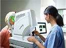
Corneal topography is indispensable in refractive surgery, but it's very useful before cataract surgery, as well. This is especially true when a surgeon intends to implant newer intraocular lenses such as multifocal and diffractive-multifocal IOLs, which demand an excellent visual system in order to yield the best results. In this article, several experts explain why they value topography highly before cataract surgery, and share tips on how to get the most out of your preop topographic exams.
Catching the Abnormal
Rockville Center
Houston
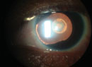
• Keratoconus and other corneal disease. When looking for keratoconus, surgeons make it a point to note any of the classic signs, such as inferior corneal steepening with skewed, asymmetric astigmatism1 and an inferior:superior ratio usually greater than 1.4 D.2 "And, every once in a while, you can have a nipple cone," says Dr. Koch, "with a lot of steepness right in the center of the cornea."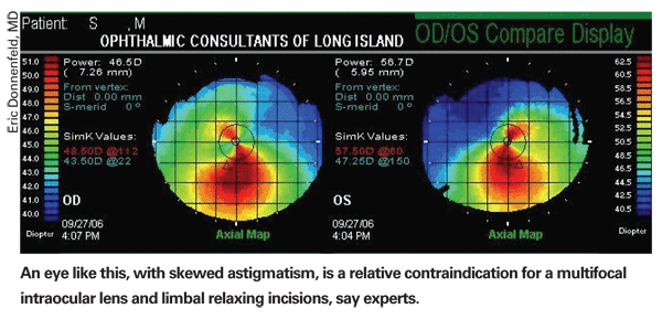
Again, surgeons note that forme fruste or frank keratoconus isn't the dire red flag it is in a LASIK patient, but it is something to talk to the patient about. "Topography's helpful in picking up the keratoconus," says
Dr. Donnenfeld notes it's good to catch skew deviation in astigmatism if you're planning to do LRIs or astigmatic keratotomy. "In skewed astigmatism, one of the wings of the 'bow tie' curves to the side, and is a warning sign of an irregular cornea," he says. "Also, such patients don't do as well with AK and LRIs because they don't respond normally, and you could end up creating a lot of irregular astigmatism with your incisions." He adds that topography is part of the diagnosis, but not the only factor. "In general, you want to make sure that the corneal topography is regular, and if it's not, you certainly would want to know something about the pachymetry to make sure you're not dealing with a frankly keratoconic eye," Dr. Donnenfeld says.
Another irregularity that can be caught on topography is the rare disease pellucid marginal degeneration, which can appear as a vertical, negative bow tie. The lower half of the bow tie may be surrounded by a claw or C-shaped elevation in corneal power. Experts say a third irregularity—epithelial, or anterior, basement membrane dystrophy—isn't as clear-cut, appearing as an amorphous irregularity similar to that caused by minor dry eye, or if the patient had just instilled some drops in his eye. "For ABMD, the biggest sign is irregular distortion or irregular mires," says
"Topography is a very useful screening tool for some corneal surface irregularities, such as epithelial basement membrane disease or Salzmann's nodular degeneration," adds Dr. Koch. "It's surprising that you'll have a patient with very few corneal epithelial changes, yet when you perform topography you see dramatic distortion or irregularity of the mires." He notes that, catching Salzmann's nodules can actually change your approach to surgery in some cases. "These patients, in my opinion, first require epithelial debridement or removal of the Salzmann's nodules. They then should be allowed to heal for two or three months," Dr. Koch says. "Then, one of two things will happen: They'll be ready for cataract surgery; or if their cataracts aren't too severe, their vision will be so improved that they'll defer the surgery. In either case, it's a win for the patient." Dr. Koch emphasizes that if a surgeon's using topography to screen for things like epithelial irregularities or Salzmann's nodules that the surgeon should refer to the placido mires first. "They provide the best information regarding distortion induced by these surface irregularities," he says. "The color-coded maps can sometimes mask these."
Dr. Hardten says topography can be helpful in another rare group of patients, as well. "I've become more concerned about patients having refractive surgery when they're 25, but not mentioning it later when they're 55 and need cataract surgery," he says. "It may be very difficult to pick this condition up on the regular slit lamp exam, so when you do your K-reading, you don't know if a 40-D K is the patient's normal K or if it's that way because he had previous refractive surgery. Corneal topography makes it a lot more obvious that it was previous surgery; if it was a myopic procedure, it will be flatter centrally. If it was a hyperopic surgery, it will be more prolate than the typical cornea."
• Corneal scars. Though you will, of course, catch a corneal scar when examining a patient that has one, Dr. Hardten says corneal topography will tell you the extent of its impact on the cornea in terms of irregularities. "Also, in a case of pterygium, topography will help you decide whether to do the cataract surgery or remove the pterygium first, in terms of which would give the patient better vision," he says. "If there's a lot of irregular astigmatism on the topography due to the pterygium, you might want to address the pterygium first."
• Ocular surface/dry eye. Dr. Hardten notes that dry eye can distort topography as well, letting you know that the surface may need to be normalized to get the best vision postop. "Sometimes dry eye can be seen more easily on topography than at the slit lamp," he says. "Because, at the slit lamp, you're probably looking primarily at the cataract rather than the ocular surface, while at the topographer you're trying to read and understand the topography."
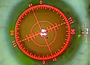
When examining topography, surgeons say it's important to be aware of the type of map you're studying, as well as the dioptric scale the map is using. "Most of us are used to looking at axial maps, which are called sagittal on some machines," explains Dr. Hardten. "Tangential maps tend to exacerbate or magnify what you'd normally see on the axial and sagittal images. However, by that magnification you can sometimes perceive abnormalities you may not have otherwise perceived on the other maps.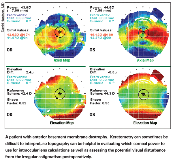
"The scale is also something to be aware of," adds Dr. Hardten. "Some topographers do auto-scaling, so the color selections and expansion of the ranges can be magnified or contracted. So, a normal cornea can look abnormal with a really expanded range of colors, and an abnormal cornea could look really normal if you've contracted down the ranges of the powers on the map … I think somewhere between a 1- and 1.5-D step between the colors usually gives a good balance of sensitivity but also avoids overcalling things."
Dr. Koch, however, likes a scale with smaller steps. "I like a scale that's relatively detailed," he says. "I'm not interested in a 1.5- or even a diopter scale. In general, I like a 0.5-D scale, because that's the level of detail I need in order to assess the impact of things such as epithelial irregularities on quality of vision."
Planning LRIs
Deciding exactly where to place LRIs is a job tailor-made for topography, surgeons say.
"With topography, and tomography [like the Pentacam or Orbscan], you can customize your incisions for the patient," says Dr. Holladay. "It's rare to have a symmetrical bow tie, and most of the time it's asymmetrical, with a bigger 'balloon' on one side than the other. You can't see this on a keratometer. When you see this on topography, you know that you can make the incision on the steeper side a little longer than on the other side. So, you can make the patient more symmetrical.
"Also, for skewed astigmatism in which the axes aren't perpendicular, you don't do the incisions on opposite sides," Dr. Holladay adds, "but place them at the tips of the astigmatism to round it out and make it more symmetrical. So, topography and tomography give you the additional information that allows you to customize the incisions for the patient."
Dr. Donnenfeld takes the topography into the OR with him when he's going to do LRIs. "I'll usually flip the topography upside down in the OR and use it as a guide for placing my LRIs," he says. Like Dr. Holladay, he also will sometimes customize the incisions for the patient. "If the two 'wings' of the astigmatism aren't 180 degrees apart and don't have tremendous skew, I'll sometimes angle my LRIs to place them at the axis of topographic cylinder rather than the axis of the cylinder on refraction."
Dr. Hardten says that the patient's irregular astigmatism may prohibit relaxing incisions. "For patients with irregular astigmatism, even though they have astigmatism in their refraction, their irregular astigmatism may not be something that's very amenable to improvement with LRIs," he says. "For those patients you may not want to do LRIs, but rather wait until after the surgery to see what happens."
Surgeons also note it's important to look for any discrepancies between astigmatism as measured by refraction and that seen on topography. "They may have 2 D of cylinder on refraction but no astigmatism on topography," says Dr. Hardten. "That's someone to be careful about doing relaxing incisions on because some of the astigmatism could be lenticular or on the back of the cornea, and you won't know exactly where until you perform the cataract surgery." Dr. Hardten will leave the astigmatism alone in a patient in whom the topographic and refractive astigmatism are significantly different. He'll treat it later with laser a few months after the surgery.
Dr. Hardten adds that the Orbscan and the Pentacam can give regional pachymetry values in the areas where you're planning your relaxing incisions. "You wouldn't want to do a 600-µm relaxing incision in someone with a 400-µm peripheral cornea," he says.
Topography also helps a surgeon mark the eye for relaxing incisions. "In people with regular astigmatism, topography allows you to get some surgical landmarks preop by seeing where the vessels around the limbus are in the image," says Dr. Holladay. "So, the day before surgery, some surgeons will nick the vessels closest to the axis on which they want to make their incisions with a 30-ga. needle. They'll do this on both sides of the meridian. When the patient comes in, there will be little red spots that tell them right where the axis is on the table. Some surgeons might use gentian violet to mark it the day before, but that mark will be gone by the time the patient comes back."
Help with Aspheric IOLs
Aspheric lenses such as the AMO Tecnis Z9000, the Alcon SN60WF and the Bausch & Lomb SofPort Advanced Optics lens have been helpful in enhancing contrast sensitivity in patients post-cataract surgery by either negating the cornea's positive spherical aberration to try to bring the visual system's spherical aberration to zero (the Tecnis and the SN60WF) or by not affecting it at all (the SofPort AO). However, if you know a patient's corneal spherical aberration, you may be able to tailor an aspheric lens for him using corneal topography and a special software program.
The program is called VOL-CT, and was developed by
When planning surgery, Dr. Donnenfeld says more information is always better. "It all comes back to the fact that, if you're putting an IOL in and the cornea is irregular," he says, "those aberrations will be transmitted through the visual system."
1. Rabinowitz YS. Corneal topography. Curr Opin Ophthalmol 1993;4:68-74.
2. Maeda N, Klyce SD, Smolek MK. Comparison of various methods for detecting keratoconus using videokeratography. Arch Ophthalmol 1995;113:7:870-4.