X-Linking for a Refractive Effect
Internationally, investigators are conducting studies to determine the efficacy of cross-linking in correcting refractive error. “It became evident when looking at the changes in the corneal shape following cross-linking for keratoconus and ectasia that it is possible to induce refractive changes,” says Michael B. Raizman, MD, who is in private practice in Boston. “Especially with some of the newer UV light devices that provide customized light profiles, it became apparent that it might be possible to control more specific changes in corneal curvature that could affect the refractive error. The next step was to consider this for normal corneas, as opposed to corneas that are ectatic from keratoconus or previous laser procedures.”
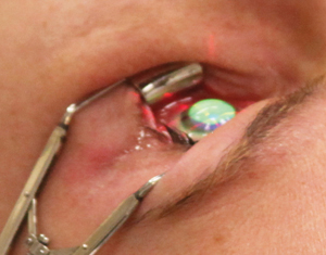
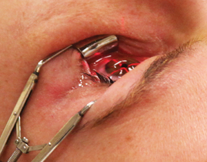
The KXL II unit by Avedro, which is used to perform photorefractive intrastromal cross-linking (PiXL), has been introduced in other countries and uses an active eye tracker to achieve a patterned delivery of UV light. PiXL has the potential to nonsurgically correct myopia and improve cataract surgery outcomes. The device delivers specific light patterns to the cornea based on a patient’s topographic data.
“PiXL has a lot of potential,” says Rajesh Rajpal, MD, who is in private practice in the Washington, D.C., area. “The concept is that, by differentially cross-linking within different parts of the cornea, the refractive error can be changed. With excimer laser treatment, we are removing tissue. PiXL affects tissue in different patterns, and because the cornea seems to change structure to some degree or at least change the curvature, ultimately, you achieve a refractive effect. This has a lot of potential, but it is still preliminary in terms of outcomes and duration of refractive result.”
|
Epithelium: On or Off?
According to Dr. Rajpal, there are continuing efforts toward better understanding whether epithelium-on or epithelium-off treatments are more effective. “I think it comes down to the type of riboflavin being used to some degree,” he says. “If we are using riboflavin that is able to be absorbed through the epithelium, then, hopefully, we are all optimistic that the effect in the stroma of the cross-linking is as good as taking the epithelium off, so we don’t have to do epithelium-off treatments. That continues to be an area where there is significant effort. Part of it just comes down to having enough treatments done to be able to analyze that data to see if the effect is as significant as the effect with epithelium-off treatments.”
Peter Hersh, MD, who is in private practice in Teaneck, N.J., agrees. “There are clinical studies going on in the United States looking at epi-on cross-linking,” he says. “The jury is still out on the efficacy of that compared to standard cross-linking. Epi-on cross-linking may be limited by a few factors, such as absorption of the incoming power by the epithelial cell layer. Also, oxygen availability, in theory, may affect epi-on outcomes. We have learned that oxygen is important in at least one of the pathways of cross-linking. Because oxygen diffusion through the epithelial layer may not be as great as when the epithelium is removed, this may be a limiting factor as well to the efficacy of transepithelial cross-linking. I do think that the ability to get riboflavin into the cornea via transepithelial technique is much improved, and this is much less of an issue than it has been in the past.”
A recent study conducted in Turkey has found that transepithelial cross-linking with prolonged preoperative riboflavin application can achieve a similar depth of effect in the stroma with less pronounced confocal microscopic changes as standard cross-linking with complete epithelial debridement.1 This study included eyes with progressive keratoconus that underwent cross-linking with the standard technique and with a transepithelial technique after prolonged riboflavin drop application for two hours. The depth of the cross-linking effect was similar in both groups (380.86 ±103.23 µm in the standard cross-linking group and 342.2 ±68.6 µm in the transepithelial cross-linking group). In the eyes that underwent standard cross-linking, anterior stromal acellular hyper-reflective honeycomb edema with posteriorly gradually decreasing reflectivity and increasing number of keratocytes and some sheets of longitudinally aligned filamentary deposits were observed on in vivo confocal microscopy. The keratocytes repopulated in a posterior-to-anterior direction. In the eyes treated with trans-epithelial cross-linking, there was less pronounced keratocyte damage, extracellular matrix hyper-reflectivity and fewer sheets of filamentary deposits at the posterior stroma.
Accelerated Cross-Linking
Accelerated cross-linking, in which the UV light is delivered at a higher intensity over shorter periods of time, is growing in popularity. Initial published reports indicate that this is a safe and effective alternative to the traditional Dresden protocol, but larger studies are needed to understand the optimal parameters. Combining accelerated treatments with pulsing of light and supplemental oxygen may be even more effective. Three multicenter studies in the United States are looking at the efficacy of accelerated cross-linking. Ultraviolet power is increased typically from 3 to 30 milliwatts, and early results are encouraging.
Dr. Rajpal has been using accelerated cross-linking in the clinical trials he is participating in. “This shortens the amount of UV exposure time,” he says. “It is easier for the patient and for the doctor, and because there is less time under the UV light, the total treatment time is shortened. The procedures have similar outcomes, so I think there is little debate that this is the direction that all treatments are going.”
X-Linking with Other Procedures
There have been many reports in the literature about LASIK Xtra, which uses cross-linking as an adjunct to standard LASIK to avoid post-LASIK ectasia and to improve refractive outcomes.
International studies have shown the benefits for patients with high myopia and high hyperopia corrections. A. John Kanellopoulos, MD, recently concluded a long-term study comparing LASIK Xtra to standard LASIK for high myopia corrections.2
|
He has also studied LASIK Xtra in patients with hyperopia and hyperopic astigmatism.3 In this study, 34 consecutive patients with hyperopia and hyperopic astigmatism elected to have bilateral topography-guided LASIK and were randomized to receive a single drop of 0.1% sodium phosphate riboflavin solution under the flap followed by a three-minute exposure of 10 mW/cm2 ultraviolet A light with the flap realigned in one eye and no intrastromal cross-linking in the contralateral eye.
At two years postop, the mean spherical equivalent refraction was -0.20 ±0.56 D and +0.20 ±0.40 D with mean cylinder of 0.65 ± 0.56 D and 0.76 ± 0.72 D and mean uncorrected distance visual acuity of 0.95 ±0.15 and 0.85 ±0.23 in the cross-linking and LASIK only groups, respectively. Eyes that underwent cross-linking demonstrated a mean regression from treatment of +0.22 ±0.31 D, whereas eyes that underwent LASIK only showed a statistically significantly greater regression of +0.72 ±0.19 D.
According to Dr. Hersh, internationally, topo-guided PRK is being performed adjunctively with cross-linking, and results are encouraging. Dr. Kanellopoulos has studied same-day topography-guided PRK followed by cross-linking, compared with cross-linking followed by topography-guided PRK six months later.4-6 Procedures performed on the same day have been found to be superior to sequential procedures for visual rehabilitation.
Dr. Kanellopoulos’ group found that 27 of 32 eyes achieved uncorrected and corrected distance visual acuity of 20/45 or better and had an improvement in uncorrected distance visual acuity and corrected distance visual acuity at last follow-up. Four eyes had topographic improvement, but no improvement in corrected distance visual acuity. Additionally, one eye required a subsequent penetrating keratoplasty, and two eyes developed grade 2 corneal haze.
Additionally, Intacs combined with cross-linking has been found to stabilize the cornea and improve corneal topography and symmetry.7 “We are continuing our own study of Intacs and cross-linking, and we find that the combination works well. Intacs provides a fairly marked topographic improvement, and there is the adjunctive effect of cross-linking and corneal stability,” Dr. Hersh says.
Also, a recent study has found that contact lens-assisted corneal cross-linking is safe and effective for performing cross-linking in corneas of less than 400 µm after epithelial abrasion, and appeared effective based on stromal demarcation line depth.8 The study included 14 eyes diagnosed with progressive keratectasia with a corneal thickness between 350 µm and 400 µm after epithelial abrasion. Mean preoperative minimum corneal thickness after epithelial abrasion was 377.2 ±14.5 µm (range: 350 to 398 µm). A significant difference in functional corneal thickness was observed intraoperatively, before epithelial abrasion, after epithelial abrasion and with contact lens and riboflavin film. Mean minimum functional corneal thickness after the contact lens was 485.1 ±15.8. Mean absolute increase in the minimum corneal thickness along with the contact lens and pre-corneal riboflavin film was 107.9 ±9.4 µm. Mean depth of the stromal demarcation line was 252.9 ±40.8 µm. No significant endothelial loss was observed, and the corneal topography was stable at the last follow-up visit.
Another procedure of interest is Keraflex, which is performed with the Vedera System by Avedro. It uses a circle or arc of microwave energy to flatten the cornea and adjunctive cross-linking to stabilize the cornea. Dr. Hersh is currently performing Keraflex in a clinical trial. Early results of his study are promising, with significant degrees of flattening seen in some patients.
The Future
Ophthalmologists are eagerly awaiting FDA approval of corneal collagen cross-linking. “Then, I think we can examine treatment optimization, which includes potential changes in the oxygen environment or oxygen delivery, possible pulsing techniques where the light is turned on and off to allow more oxygen diffusion, and obviously the various alternate epithelial approaches,” Dr. Hersh says. “All of these need to be refined. I think the next big leap is going to be topography-guided cross-linking. It really makes sense as a treatment of irregular corneas, and I think it is an exciting potential modality for the treatment of low degrees of refractive errors. We’d also love to get topography-guided LASIK available in the United States so that we can work on combining that technique with cross-linking as well.”
He notes that cross-linking is also being used overseas as a treatment of infectious keratitis. “People are trying to optimize protocols to either use cross-linking adjunctively with current antibiotics or potentially to use cross-linking alone for some kinds of infections,” he explains.
Dr. Raizman adds that another exciting new area is cross-linking using UV light and alternative molecules other than riboflavin or even cross-linking without the use of UV light. “Perhaps there are better ways to cross-link. It is a rapidly evolving area,” he says. REVIEW
1. acar b, utine c, ozturk v, acar s, ciftci f. can the effect of transepithelial corneal cross-linking be improved by increasing the duration of topical riboflavin application? an in vivo confocal microscopy study. eye contact lens 2014 may 28. [epub ahead of print].
2. kanellopoulos aj, asimellis g. comparative epithelial topography and thickness changes following femtosecond-assisted high myopic lasik with versus without prophylactic higher-fluence collagen cross-linking. cornea 2014 (accepted for publication).
3. kanellopoulos aj, kahn j. topography-guided hyperopic lasik with and without high irradiance collagen cross-linking: initial comparative clinical findings in a contralateral eye study of 34 consecutive patients. j refract surg 2012;28(11 suppl):s837-s840.
4. kanellopoulos aj. comparison of sequential vs same-day simultaneous collagen cross-linking and topography-guided prk for treatment of keratoconus. j refract surg 2009;25:s812-818.
5. krueger rr, kanellopoulos aj. stability of simultaneous topography-guided photorefractive keratectomy and riboflavin/uva crosslinking for progressive keratoconus: case reports. j refract surg 2010;26:s827-832.
6. kanellopoulos aj, binder ps. management of corneal ectasia after lasik with combined, same-day, topography-guided partial transepithelial prk and collagen cross-linking: the athens protocol. j refract surg 2011;27:323-331.
7. hovakimyan m, guthoff rf, stachs o. collagen crosslinking: current status and future directions. j ophthalmol 2012;epub 2012 jan 12.
8. jacob s, kumar da, agarwal a, basu s, sinha p, agarwal a. contact lens-assisted collagen cross-linking (cacxl): a new technique for cross-linking thin corneas. j refract surg 2014;30(6):366-372.


