A 73-year-old Caucasian woman with gradual onset of blurred vision and pressure sensation in her left eye for two months presented for ophthalmic evaluation. She had a worsening of chronic headaches and new onset of floaters in the left eye, as well. She initially described her symptoms to her primary care provider who referred her to an optometrist. The optometrist saw a suspicious choroidal lesion on dilated examination of the left eye and referred her to a retinal specialist, who subsequently referred the patient to an ocular oncology specialist to rule out choroidal malignant melanoma.
Medical History
Past ocular history included cataract extraction with placement of an intraocular lens in the right eye one year prior and primary
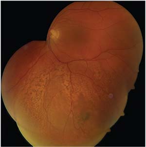 |
| Figure 1. Montage color fundus photo of the left eye at the patient’s first visit to the ocular oncology clinic. An ill-defined, 16 x 14-mm amelanotic mass is present inferior to the inferior arcade, disrupting the retinal pigment epithelium. |
Examination
On examination, visual acuity was 20/20 in each eye. Pupils were symmetric and without afferent pupillary defect. Intraocular pressures were 13 mmHg in the right eye and 14 mmHg in the left. Extraocular movements were full bilaterally and confrontation visual fields were full in both eyes.
On examination of the anterior segment, the right eye revealed a posterior chamber intraocular lens, and the left eye had mild nuclear sclerosis and anterior cortical changes. Dilated fundus examination of the right eye demonstrated normal macula, disc, vessels and periphery, with a cup/disc ratio of 0.6 and evident posterior vitreous detachment. The left fundus was similar, with a normal macula and a cup/disc ratio of 0.6. However, in the inferior quadrant, there was an ill-defined yellow mass deep in the retina, measuring 16 x 14 mm in diameter and associated with shallow subretinal fluid, surrounding retinal pigment epithelial alterations and a large RPE detachment (Figure 1).
Click here for diagnosis, workup and discussion.


