He explains that by making a LASIK flap and removing tissue, some important anterior aspects of the corneal structure are weakened, and corneal rigidity may decrease. Dr. Hersh cites London researcher and ophthalmologist John Marshall, PhD, who has estimated that LASIK may weaken the cornea by 15 percent to 25 percent. “In the vast majority of cases, this doesn’t lead to any clinical problem, and it certainly doesn’t lead to corneal ectasia in the vast majority of cases,” Dr. Hersh says. “Cross-linking, on the other hand, has been shown to reliably strengthen the cornea. So, the concept of combining the procedures both from a safety and efficacy point of view is of great interest.”
Dr. Hersh notes that the combined procedure, dubbed LASIK Xtra, can be viewed as an extra safety element in patients who may have some theoretically greater risk: those who have thinner corneas and those who have high degrees of correction. “Strengthening the cornea with concurrent cross-linking may be beneficial in the longer-term stability of the refractive result and ultimate predictability of the procedure, he says. “Clinical studies are showing encouraging results, and further clinical work should continue to elucidate the advantages and disadvantages of the procedure.”
Who Should Have LASIK Xtra?
Although LASIK Xtra is not approved in the United States, surgeons outside of the United States have been performing the procedure, and some believe that cross-linking should be performed on all LASIK patients.
|
A. John Kanellopoulos, MD, who is in private practice in Athens, Greece, and in New York City, employs cross-linking in all hyperopic LASIK cases. He also uses it in all young myopic patients over 6 D and under 30 years of age and in all patients with myopic astigmatism when the difference in astigmatism between the two eyes is more than 0.75 D. “For example, if one eye is 1 D and the other eye is 2 D, in my protocol, this is a reason for that patient to undergo additive collagen cross-linking regardless of age,” Dr. Kanellopoulos says.
International studies have shown the benefits for patients with high myopia and high hyperopia corrections. Dr. Kanellopoulos recently concluded a long-term study comparing LASIK Xtra to standard LASIK for high myopia corrections.1 “The results were compelling in favor of the LASIK Xtra cases as far as refraction accuracy and stability,” he says.
In this study, 65 eyes underwent LASIK Xtra and 75 eyes underwent LASIK alone. In the LASIK Xtra group, the mean patient age at the time of the procedure was 27.5 ±6.1 years (range: 19 to 39). Preoperatively, the mean refractive error was -6.60 ±2.02 D of sphere (range: -2.50 to -11.50), -1.35 ±1.24 D of cylinder (range: 0 to -5 D), and -6.75 ±1.75 D of manifest refractive spherical equivalent (range: -2.50 to -11.50).
In the LASIK only group, the mean preoperative refractive error was -5.14 ±1.74 D of sphere (range: -2.50 to -9.50), -0.85 ±0.75 D of cylinder (range: 0.00 to -3.50), and -5.33 ±2.34 D of manifest refractive spherical equivalent (range: -2.50 to -9.50). Mean central corneal thickness was 553.51 ±19.11 µm (range: 503 to 592) preoperatively and 454.34 ±19.98 µm (range: 422 to 515) one year postoperatively.
In the LASIK Xtra group, 90.8 percent of eyes had a postoperative uncorrected distance visual acuity of 20/20 (1.0 decimal) or better, and 95.4 percent had a UDVA of 20/25 (0.8 decimal) or better. In the LASIK only group, 85.3 percent of the eyes had a postoperative UDVA of better than 20/20 (1.0 decimal), and 89.3 percent had better than 20/25 (0.8 decimal). The differences between the two groups at the 20/20 and the 20/25 levels were statistically significant (p=0.045 and 0.039, respectively).
When comparing the preoperative corrected distance visual acuity versus postoperative uncorrected distance visual acuity, in the LASIK Xtra group, 35 percent of the eyes were unchanged, 57 percent gained one Snellen line, and 8 percent gained two or more Snellen lines. No eye lost any lines. In the LASIK only group, 35 percent of the eyes were unchanged, 59 percent gained one Snellen line, and 5 percent gained two or more lines. Only 2 percent (one eye) lost one line.
In the LASIK Xtra group, 85 percent of eyes had a postoperative spherical equivalent refraction between -0.5 and 0 D, compared with 83 percent in the LASIK only group. Additionally, the LASIK Xtra group had a mean preoperative cylinder of -1.39 D, while the LASIK only group had a mean preoperative cylinder of -0.86 D. Despite this, postoperatively, 92 percent of the eyes in the LASIK Xtra group had less than 0.25 D of refractive astigmatism, compared with 94 percent in the LASIK only group.
The refractive stability is demonstrated by the manifest refractive spherical equivalent correction as seen during the one-, three-, six- and 12-month postoperative visits. One-year postoperative mean manifest refractive spherical equivalent minus the one-month baseline was -0.24 ±0.09 D in the LASIK Xtra group and -0.27 ±0.09 D in the LASIK only group. These results indicate a reduced refractive shift in the LASIK Xtra group compared to the LASIK only group. The keratometric stability is demonstrated by the K-flat and K-steep average values up to the 12-month postoperative visit. The results indicate an increased keratometric stability in the LASIK Xtra group (one year at +0.03 D in the flat and +0.05 D in the steep in comparison to one month baseline), when compared to the LASIK only group (+0.57 D and +0.54 D, respectively).
“These data suggest that LASIK Xtra is a refractive stabilizer in high myopia, presumably through its biomechanical stabilization effect,” Dr. Kanellopoulos says.
|
Preoperatively, the mean refractive spherical equivalent was +3.15 ±1.46 D and +3.40 ±1.78 D with a mean cylinder of 1.20 ±1.18 D and 1.40 ±1.80 D and mean uncorrected distance visual acuity (decimal) of 0.1 ±0.26 and 0.1 ±0.25 in the cross-linking and LASIK only groups, respectively. At two years postoperatively, the mean spherical equivalent refraction was -0.20 ±0.56 D and +0.20 ±0.40 D with mean cylinder of 0.65 ±0.56 D and 0.76 ±0.72 D and mean uncorrected distance visual acuity of 0.95 ±0.15 and 0.85 ±0.23 in the cross-linking and LASIK only groups, respectively. Eyes that underwent cross-linking demonstrated a mean regression from treatment of +0.22 ±0.31 D, whereas eyes that underwent LASIK only showed a statistically significant greater regression of +0.72 ±0.19 D (p=0.0001).
“Topography-guided hyperopic LASIK with or without intrastromal cross-linking is safe and effective, with greater long-term efficacy (less regression) in eyes with cross-linking. Our data suggest that the regression seen with hyperopic LASIK may be related to biomechanical changes in corneal shape over time,” Dr. Kanellopoulos says.
U.S. Trial
In the United States, a five-center clinical trial evaluating LASIK Xtra has just gotten under way. Dr. Rajpal was the first refractive surgeon to perform LASIK Xtra in the United States. “Because we are going through the Food and Drug Administration approval process, we will ultimately have data that will show whether there is a difference between regular LASIK and LASIK Xtra,” he says. “Hyperopic treatments are a good way to do that because we may be able to demonstrate stability or lack of regression in a reasonable time period. Because the rate of ectasia is so low, if the study was done to determine only whether there was a difference in the rate of ectasia, it would be difficult to show statistical significance unless we treated hundreds of thousands of patients. That’s why the hyperopic study was started in this country.”
Downsides of LASIK Xtra
“We have very good data internationally showing that performing cross-linking doesn’t have an effect on refractive outcomes, which has been one of the concerns,” Dr. Rajpal says. “Does strengthening the cornea change what you are achieving in terms of visual outcomes? While there may be a need for some adjustment in the nomogram for some patients, internationally, the data that we have seen has demonstrated that there is not really a need for adjustment. So it seems to be having some effect, and the refractive outcome does not seem to be negatively affected in any way. I feel if we can get FDA approval for this in the United States, and hyperopic LASIK may be the pathway for it, that would be a useful option for surgeons who want to offer this to their patients. I think most doctors will choose to start off with high-risk patients. Then, if they feel that it is providing a benefit to those patients, they will be much more likely to start offering it to all patients.”
He notes that there is likely no downside to the treatment other than adding a little bit of time to the procedure by applying the riboflavin and then the UV light. “That adds about three to four minutes to the total treatment,” he says. “Cost may be the only other downside. The study we are doing is using a pulsed UV light, as there is evidence that the cross-linking effect is greater when the light is pulsed. When ectasia occurs following refractive surgery, it can be very difficult to treat. So, if we can provide a procedure for our patients that reduces that risk and doesn’t add risk or significant cost, then I think it does make sense theoretically, and we are in the process of getting more data to show that.”
According to Dr. Kanellopoulos, another downside is the possibility of infection, which can result from contaminated riboflavin solution or over cross-linking. “This is the reason we use single-use containers for riboflavin and use a strictly sterile environment in a similar fashion as we do with a routine LASIK procedure,” he says. “High-fluence cross-linking is used to avoid extensive time under the UV source and a possibility for inadvertent contamination from the operating room or the health-care staff.”
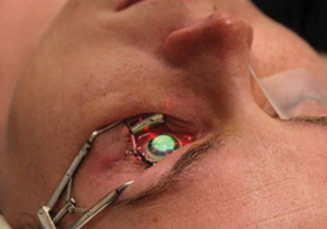
Dr. Kanellopoulos notes that there is no significant learning curve when combining the two procedures. “The basic principle is to soak the underlying stroma after the end of the ablation with riboflavin, and we have chosen the one that is diluted in saline,” he says. “It is important to avoid having the riboflavin come into contact with the flap. This is why, when I pull the flap off the cornea following the femtosecond laser flap creation, I try to fold it onto itself, thus reducing its dehydration and secondarily protecting it from any riboflavin spilled over from its instillation at the end of the ablation. The soaking takes 60 seconds, and following that, the flap is repositioned in place, and the interface is rinsed copiously in order to remove any residual riboflavin and to minimize the amount of riboflavin that jumps into the flap. After repositioning the flap, I use a Johnston applanator to iron out the central part of the cornea. I use BSS to lubricate the surface. At the end of the case, I place a bandage contact lens, which I remove the next morning.” REVIEW
1. Kanellopoulos AJ, Asimellis G. Comparative epithelial topography and thickness changes following femtosecond-assisted high myopic LASIK with versus without prophylactic higher-fluence collagen cross-linking. Cornea 2014 (accepted for publication).
2. Kanellopoulos AJ, Kahn J. Topography-guided hyperopic LASIK with and without high irradiance collagen cross-linking: Initial comparative clinical findings in a contralateral eye study of 34 consecutive patients. J Refract Surg 2012;28(11 Suppl):S837-S840.