|
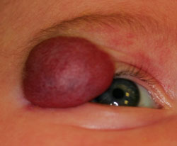
These lesions can be superficial (50 to 60 percent), subcutaneous (15 percent), or mixed (25 to 35 percent). Superficial hemangiomas, also known as “strawberry angiomas” are generally well-demarcated, red-purple, smooth lesions with a firm consistency (See Figure 1). Deep hemangiomas, also known as “cavernous hemangiomas,” are well-defined protruding masses covered by normal-appearing skin with a deeper blue-purple color (See Figure 2). Deep hemangiomas can mimic subcutaneous lymphangiomas or orbital dermoids, and neuroimaging is often necessary to determine the lesion’s extent. Hemangiomas can also be classified as localized or segmental.
Systemic, Ocular Complications
Anatomic location and size dictate potential morbidity from these otherwise benign lesions. Any of the skin lesions may be disfiguring and potentially leave fibrovascular scarring after involution. Large facial hemangiomas can be associated with PHACES syndrome (posterior fossa malformations, hemangioma, arterial anomalies, cardiac defects, eye abnormalities, sternal clefting). Lower face and neck hemangiomas have been associated with concomitant airway hemangiomas that may cause significant bleeding during anesthesia. Lesions overlying the lumbosacral spine have been associated with tethered cords and other genitourinary abnormalities.
|
Periorbital and orbital hemangiomas can be associated with vision loss from several etiologies. Eyelid lesions can cause ptosis and occlusive amblyopia. They may also be associated with induced astigmatic error causing refractive amblyopia that may not resolve with hemangioma treatment alone. Orbital hemangiomas are associated with proptosis and secondary exposure keratopathy, ocular motility restrictions and optic nerve compromise if the posterior orbit is involved. With the new appreciation that these lesions reach full size by 5 months, early referral and evaluation for ocular complications is encouraged for early treatment consideration.
Treatment Options
Treatment is indicated to prevent life-threatening complications (with airway or liver involvement), to prevent functional impairment (vision loss/amblyopia), and to prevent or improve ulceration and pain with potential long-term scarring and disfigurement.3 Observation alone is a very reasonable choice for lesions that do not seem to be interfering with vision, causing significant deformity or threatening life.
• Corticosteroids. Until recently, both intralesional and systemic corticosteroids have been considered the mainstay of treatment when observation combined with amblyopia therapy fails. Systemic steroids of varying doses can be used with a reasonable response in many cases. Systemic steroids carry potentially significant risks, especially in the infant population, including adrenal suppression, growth retardation and immunosuppression. Possible local eye complications from systemic steroids include steroid induced glaucoma and cataract formation. Intralesional injections of steroids are also an option, but have also been reported to have side effects of localized fat atrophy, cutaneous pigmentary changes, and rarely, central retinal artery occlusion. Steroid therapy needs to be closely monitored by the pediatrician and the ophthalmologist throughout the treatment course.
• Laser. Pulse-dye laser has been used for hemangiomas with some improvement. Generally this has been advocated during the early proliferative phase or the late regression phases when the lesion is flatter.
• Surgical excision. Well-circumscribed superficial lesions can be surgically excised with good results. Surgery tends to more successful in the early phases when the lesions are smaller and more confined. Because these are vascular tumors, intraoperative bleeding is a risk factor. Surgical excision in the late involuted phases may be of value to remove unsightly residual lesions.
• Oral propranolol. Propranolol is a non-selective beta-blocker used in children for several decades for cardiac, neurologic and endocrine diseases with a good safety and tolerance record. In 2008, Christine Leaute-Labreze, MD, and colleagues described two cases of resolution of capillary hemangiomas when the patients were started on oral propranolol for cardiac disease. They subsequently treated nine additional children without cardiac disease with oral propranolol for capillary hemangioma. All patients had changes in the hemangioma within 24 hours and had resolution of the lesions over the course of treatment without significant systemic side effects from the medication.4
|
Since this initial report, multiple reports have emerged using oral propranolol as a primary treatment with excellent results. These reports have also helped to define a safe and effective protocol for its use. Reported side effects from treatment include bradycardia, hypotension, hypoglycemia, allergic reaction to the medication and gastrointestinal upset. Unfortunately, not all lesions respond. Some regress within weeks of starting propranolol; others continue to grow unfettered.
Pre-treatment clearance by the pediatrician for baseline vital signs including heart rate and blood pressure and finger stick glucose testing are necessary. Electrocardiogram is indicated prior to starting treatment and if found to be irregular, then subsequent cardiology evaluation and echocardiogram are indicated.
Current dose recommendations are to initiate treatment at 0.5 mg/kg/day divided in two to three doses and to taper up to 2.0 mg/kg/day divided into two to three doses over the course of the first several days of treatment.5-7 Many providers initiate therapy in the inpatient setting to closely monitor for cardiac and respiratory side effects, followed by periodic outpatient evaluation by the primary care physician. Kathryn M. Haider, MD, and colleagues recently reported safe initiation of treatment entirely in the outpatient setting with close monitoring by the parents and the pediatrician.7 Parents must be instructed to administer the medication with meals and avoid pre-bedtime doses to avoid nocturnal hypoglycemia.
Treatment is continued throughout the proliferative phase and can be tapered and stopped when sufficient regression has been achieved. In their study, Patrizia Vassallo, MD, and colleagues reported complete resolution of hemangiomas in four months of treatment in patients under 1 year old.6 In their older patients, longer treatment did not yield greater results, likely because these lesions were already out of the proliferative phase. Recurrence of the lesions requiring restarting therapy was reported infrequently.
• Topical timolol. Recently, investigators have begun to study the effects of topical beta blockers on small, well-defined hemangiomas.8,9 Timolol maleate is a non-selective beta blocker similar to propranolol and historically used to treat glaucoma. Several authors have shown a regression response in hemangiomas treated with topical solution. Christopher Chambers, MD, and colleagues showed good response using timolol maleate gel 0.25% twice a day for superficial and mixed type hemangiomas. The one deep lesion in their study did not respond to topical therapy. They reported no adverse ocular or systemic side effects.
Infantile hemangiomas are benign lesions histopathologically, but may carry significant systemic and ocular morbidity and potential mortality. Early intervention for these high-risk lesions should be taken to avoid irreversible complications and also to provide improved cosmetic results as these children get older. PHACES syndrome should be considered in patients with segmental lesions over 5 cm, and workup should be completed before considering systemic treatment. Treatment considerations include systemic propranolol, surgical excision, systemic or injected corticosteroids, pulse-dye laser and topical timolol. Propranolol therapy has been shown to be safe and effective in reducing the size of these lesions and promoting rapid and permanent regression. Topical beta-blocker therapy is emerging as promising therapy for smaller, less aggressive lesions. REVIEW
Dr. Salvin practices in the Division of Ophthalmology at Nemours/A.I. DuPont Hospital for Children, and the Department of Ophthalmology and Pediatrics at Jefferson Medical College/Wills Eye Institute.
1. Chang LC, Haggstrom AN, Drolet BA, et. al. Growth Characteristics of Infantile Hemangiomas: Implications for Management. Pediatrics 2008;122:360-367.
2. Neri I, Balestri R, Patrizi, A. Hemangiomas: New insight and medical treatment. Dermatologic Therapy 2012;25: 320-334.
3. Ni N, Gua S, Langer P. Current concepts in the management or Periocular infantile (capillary) hemangioma. Curr Opin Ophthalmol 2011;22:419-425.
4. Leaute-Labreze C, Dumas de la Roque E, Hubiche T, et al. Propranolol for severe hemangiomas of infancy. N Engl J Med 2008;358:2649-2651.
5. Missoi T, Lueder GT, Gilbertson K, et al. Oral propranolol for the treatment of Periocular infantile hemangioma. Arch Ophthalmol 2011;129:899-903.
6. Vassallo P, Forte R, Di Mezza, A, et al. Treatment of infantile capillary hemangioma of the eyelid with systemic Propranolol. Am J Ophthalmol 2013;155:165-170.
7. Haider KM, Plager DA, Neely DE, et al. Outpatient treatment of Periocular infantile hemangiomas with oral Propranolol. J AAPOS 2010;14:251-256.
8. Chambers CB, Katowitz, WR, Katowitz, JA, et al. A controlled study of topical 0.25% Timolol maleate gel in the treatment of cutaneous infantile capillary hemangioma. Ophthal Plast Reconstr Surg 2012;28:103-106.
9. Chakkittakandiyil A, Phillips R, Frieden IJ, et al. Timolol maleate 0.5 percent or 0.1 percent gel-forming solution for infantile hemangioma: A retrospective, multicenter, cohort study. Pediatric Dermatology 2012;29:28-31.


