Ocular trauma, although not an everyday encounter for many ophthalmologists, is a serious problem for our health system and economy. An estimated 2.4 million eye injuries annually result in more than a billion dollars a year in total costs to society. (According to the United States Eye Injury Registry, 95 percent of ocular trauma injuries occur in males, most of whom are under 30 years of age; sadly, most of these injuries are preventable.)
In general, the more serious types of ocular trauma—such as ruptured globes and corneal lacerations requiring surgical reconstruction—are less frequently seen by most ophthalmologists. Here, three surgeons with extensive experience discuss their approaches to managing these kinds of trauma.
Initial Presentation
When a patient who has suffered ocular trauma is first seen, the initial objective must be to determine the nature and extent of the injury. (Of course, every injury is unique, so any step-by-step guidelines must be adapted to the situation at hand.)
Franco M. Recchia, MD, associate professor of ophthalmology and visual sciences and chief of the Retina Division at the Vanderbilt Eye Institute in
"You want to make sure the patient doesn't have any other occult injuries that could be more life-threatening than the ocular injury," he explains. "Then, a history is helpful both for clinical management and for documentation in case of legal action down the road. Was it a high-velocity injury? Is there a possibility of penetration with a retained foreign body? Was there assault? Were drugs involved? In the case of a motor vehicle collision, what were the details? Once you've made a global or systemic assessment and taken a history of the circumstances surrounding the injury, your examination should begin."
The surgeons list a number of key steps to take during your examination:
• Check visual acuity. Dr. Recchia says this should be the first order of business in the examination. "Whether the patient has light perception or no light perception is very important with respect to prognosis, especially if the globe turns out to be open," he notes.
• Determine what is injured. M. 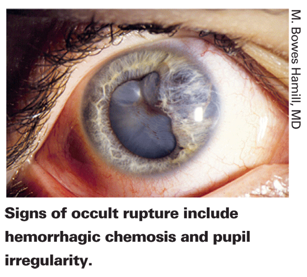
"It's important to do a complete eight-point ophthalmic evaluation," he continues. "One key issue is whether there are intraocular injuries. If you can see through the cornea and there's nothing in the anterior chamber and you can do a complete examination of the angle, iris, lens, vitreous and the posterior segment with the slit lamp and an indirect ophthalmoscope, you can be pretty sure there's no occult damage. On the other hand, if the patient has a corneal laceration and you're not sure whether a foreign object entered the eye, or the eye is full of blood obscuring your view, you'll need to do a CT scan."
• Determine whether or not the globe has been ruptured. "Sometimes a ruptured globe is fairly obvious because the globe is misshapen or contents are extruding," notes Dr. Recchia. "However, a rupture in the globe can also be fairly subtle, so you need to check for other clinical signs that suggest that the eye is open. Such signs would include the IOP in the traumatized eye being significantly lower than the fellow eye; if the pupil is a different size from the other eye or is misshapen in some way, suggesting that the iris may be plugging a peripheral wound; if the anterior chamber is shallower or deeper than the fellow eye; and if there is significant chemosis—usually 360 degrees and usually hemorrhagic—suggesting that there may be bleeding through a scleral rupture."
Management is entirely different depending on whether or not the globe is ruptured. "Closed-globe injuries usually involve blunt force to the face or eye," Dr. Recchia explains. "If the globe is closed, we follow an algorithm of checking to see what the injuries are. Are there any associated injuries such as orbital fractures or lacerations that need attention? Does the patient require antibiotics? This may be the case if there's an orbital fracture. Does the eye have a hyphema? Of course, we also want to examine the retina to make sure there's no obvious retinal tear or detachment. If the eye has a traumatic cataract that blocks the view, a gentle ultrasound through closed lids can help to evaluate for retinal detachment or tear. It's also important to look for any sign of traumatic optic neuropathy, which may manifest as acute swelling or hemorrhaging within the optic nerve."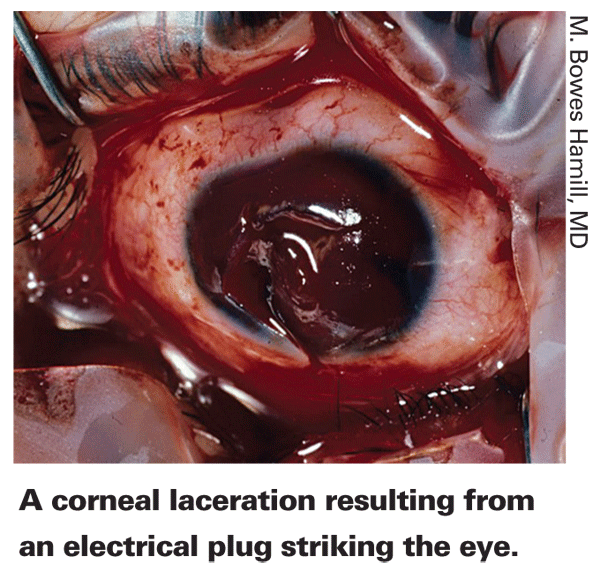
Dr. Recchia notes that an open globe injury involving a full-thickness wound, in either the cornea or sclera or both, is an emergency that requires prompt evaluation and may require surgical wound closure. "If you suspect a rupture, protect the eye from any pressure that could cause further extrusion of intraocular contents," he says. "Place a hard shield on the eye, keep the patient upright and tell him not to rub the eye. Also, make sure the patient doesn't consume any food or fluids in case surgery has to be done immediately. Confirm that the patient is up to date with his tetanus shots. Then, get the patient to the OR as promptly as possible."
Dr. Hamill notes that you'll also need to arrange to have the tools you require available at the hospital or emergency room you're sending the patient to. "If it's a bad corneal laceration and you'll need patch material, arrange to get that," he says. "If you need a vitreoretinal colleague to assist, call him and make the arrangements."
Orbital Concerns
Peter J. Dolman, MD, FRCSC, clinical professor in the department of ophthalmology and visual sciences at the
"I look for six types of injury when faced with ocular trauma," says Dr. Dolman. "In order of urgency, these are: injury to the globe itself; compartment syndrome; optic nerve injury; bony injury; foreign bodies in the orbit; and injury to the eyelids.
• Compartment syndrome. "Once you've addressed the condition of the globe itself, you should look for signs of compartment syndrome, which refers to a sudden rise in pressure in the orbital tissues," he explains. "The orbit is a closed space, and compartment syndrome can arise if anything accumulates within that space—pus produced by an infection, sudden bleeding inside the orbit, or air trapped inside the orbit. The resulting increase in pressure can damage the optic nerve or raise pressure inside the eye, which can lead to interruption in the blood supply to the eye and loss of vision.
"Signs of compartment syndrome include the eye being prominent, i.e., pushed forward; tense eyelids; and/or restricted globe movement," he continues. "You can also feel tension if you gently press on the globe. It's important to look for these signs because compartment syndrome can cause permanent vision loss within half an hour if the pressure is high enough—but it's reversible in many cases if you take quick action."
"If you believe you're dealing with acute compartment syndrome, you can perform a canthotomy and inferior cantholysis in the office, separating the lower lid from its tight insertion into the canthus at the lateral aspect of the lid," he says. "In most cases that will relieve the pressure on the optic nerve and potentially reverse any vision loss. You should arrange for a subsequent CT scan to determine what the underlying cause of the compartment syndrome was so that any need for draining can be addressed. Any minor trauma to the cornea or lids can be repaired later."
Dr. Dolman adds that the patient doesn't necessarily need to be hospitalized if this approach releases the pressure and vision returns. "But the patient probably should be evaluated by an orbital surgeon or oculoplastic surgeon if one is available," he says, "and a CT scan should be done as soon as possible."
• Optic nerve compression. Dr. Dolman says the third thing the ophthalmologist needs to look for is optic nerve compression, or traumatic optic neuropathy, an anatomical disruption of optic nerve fibers that can be caused by penetrating orbital trauma, bone fragments in the optic canal or hematomas in the nerve sheath. "Signs of traumatic neuropathy include loss of vision that can't be accounted for by other factors such as direct injury to the globe," he explains. 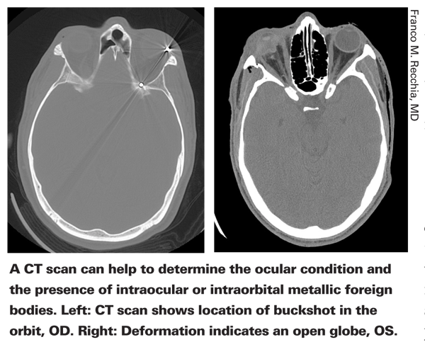
"These patients usually present with an afferent pupil defect, a drop in central vision and poor color vision," he continues. "
Less-Urgent Orbital Concerns
Dr. Dolman notes that the remaining items on his list—bony injuries, foreign bodies in the orbit and injury to the eyelids—can be observed for several days before taking action. "They can be documented by clinical examination and CT scan," he notes. "A referral to a plastic surgeon or oculoplastic surgeon can wait several days to a week."
• Bony injuries. Dr. Dolman explains that bony fractures within the orbit usually occur in the floor of the orbit or the medial wall. "Those are the two weak spots of the orbital walls," he says. Common clinical signs of a blowout fracture include a history of blunt trauma to the eye; an eye that is enophthalmic (appearing sunken); numbness in the cheek (because the nerve that supplies sensation to the cheek runs through the floor of the orbit where the fracture occurs); and an eye that doesn't elevate upwards or downwards well, leading to binocular double vision (because a muscle is entrapped in a bone fragment). "A blowout fracture is typically confirmed with a CT scan," he notes. "Both axial and coronal scans should be performed."
• Foreign body in the orbit. "Suspicion of this usually arises because of the circumstances surrounding the injury," he says. "For example, if the patient was in a car accident involving broken glass or was hit with a beer bottle, glass fragments could be embedded in the orbit. If the patient was riding a bike and went into a tree, there could be wood fragments in the orbit. You should be especially suspicious if there are perforating wounds through the eyelids or globe." Dr. Dolman says a CT scan or MRI can help to rule out the presence of a foreign object in the orbit; in most cases this type of problem would be managed by an oculoplastic or orbital surgeon.
"Management can be delayed in many cases," he notes. "If the foreign object is a small metal fragment such as a BB pellet, you may be able to leave it alone as long as it isn't injuring the globe or pressing on the optic nerve. However, any material that's close to the surface or impinging on structures, or is organic or copper, should be removed quickly to prevent inflammation or infection."
• Lid injury. The last thing on Dr. Dolman's list is eyelid injuries. "Many ophthalmologists can manage simple eyelid injuries," he notes. "You can always choose to refer more complicated cases. In most instances, as long as the eye is kept moist and protected, an eyelid injury can be repaired several days later."
In the Hospital
When a patient's wounds require going to the hospital for immediate surgery, surgeons offer these suggestions:
• Start an IV. "Once you're in a hospital or emergency room setting, start an IV," says Dr. Recchia. "Some people suggest giving the patient prophylactic intravenous antibiotics. That's debatable because a lot of those antibiotics don't have adequate penetration into the eye—although there is good evidence that fourth-generation fluoroquinolones such as moxifloxacin achieve sufficient levels within the eye. As a retinal surgeon, I have a low threshold for resorting to intracameral or intravitreal antibiotics if there's any possibility that the eye has been seeded with organisms, unless I have reason to suspect choroidal or retinal detachment. Remember that the risk of endophthalmitis is much higher in cases of dirty wounds, cataract or penetration of the lens capsule."
Dr. Recchia adds that even if you don't give intravenous antibiotics it's still important to insert the IV. "You'll need it for hydration during surgery," he says. "You'll also want to limit nausea and vomiting, which can strain the eye. Some type of anti-emetic such as compazine or phenergan can be given intravenously, along with something for pain control."
• Do any needed ancillary studies. "Generally, that means a CT scan or X-ray," says Dr. Hamill. "Sometimes you'll do blood studies if you have a patient with a hyphema and you're looking for sickle cell anemia. But the vast majority of cases of possible intraocular foreign bodies call for radiography."
Dr. Recchia agrees. "A scan can rule out the presence of an intraocular foreign body, even though the history may imply there isn't any," he says. "I've learned never to trust the history completely, especially in trauma cases."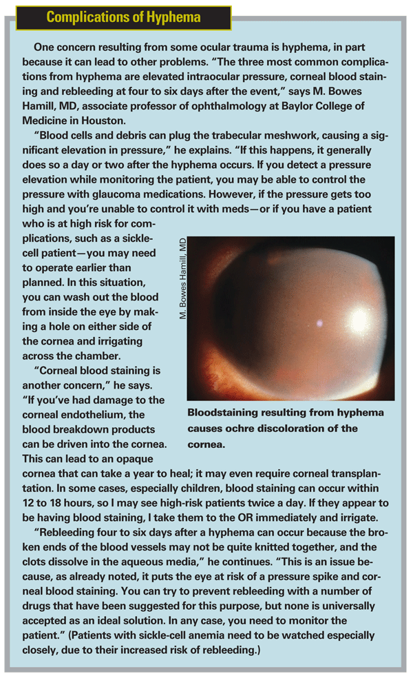
Once surgery has begun:
• Manage prolapsed tissue. "If the prolapsed tissue consists of vitreous or lens fragments, you can cut them off at the surface of the wound," notes Dr. Hamill. "However, if it's iris or other viable tissue, I'd urge you NOT to blindly excise it. In the old days we were taught to excise anything that had been prolapsed out of the eye for four or five hours. But even iris that's nonfunctional still serves a purpose in terms of light blockage. In the acute phase, reposit it back inside the eye unless it's grossly necrotic or infected.
"If the iris has been prolapsed for several days it may have a covering of epithelium," he adds. "In this situation the prolapsed tissue can be treated with a cryoprobe—freeze/thaw times two—and the epithelium can be peeled off. The treated iris can then be reposited. You can worry about doing reconstruction after the eye has been closed."
• Close the eye. Dr. Hamill says the primary concern here should be to restore normal anatomy. "It's not enough just to get the eye closed and watertight," he notes." If you don't get the anatomy right, you're not going to restore function.
"The first step when closing the eye is to figure out where your landmarks are," he explains. "Identify the places that can easily be confirmed as belonging together and stitch those together first. For example, if a laceration involves the limbus, you can put your first stitch at that easily identifiable location. Then look for jigsaw puzzle pieces that clearly match and put your next stitches there, treating each separate little arm of the laceration individually. Eventually you'll get the torn tissue correctly repositioned and restore the normal external anatomy. In most cases, tissue that seems to be missing will turn out to be just folded under."
Dr. Recchia says the goal is to explore the extent of the wound and then close it with as little collateral damage as possible. "If it's a purely corneal wound, we generally use 10-0 nylon sutures to close the wound," he says. "We re-form the anterior chamber and then bury the knots. If it's a corneal-scleral wound, we use 10-0 nylon for the cornea, 9-0 nylon at the limbus and 8-0 nylon for the sclera."
Dr. Hamill says that once the cornea is closed, if the laceration goes back into the sclera, he starts exploring to see how far back it goes. "Up to this point, the periuveal tissues are holding the intraocular tissues in place," he notes. "For that reason you don't want to expose everything at once; you could end up with a deflated eye. I recommend using a 'close as you go' method. Open the periorbita to expose a little of the scleral laceration and close what you find; then open a little more and close what you find. Repeat this until you reach the end of the laceration—or until the laceration goes so far back that you can't get to it. Once you've accomplished that, you'll have a closed, anatomically restored globe and you're well on your way to restoring function."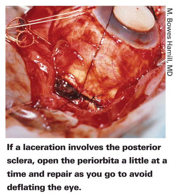
Dr. Recchia says that if a scleral wound goes so far posteriorly that you can't pass sutures, it's safer to leave the most posterior part of the wound unsutured. "Trying to suture it could put pressure on the globe, causing intraocular contents to extrude," he explains. "For that reason, we sometimes leave very posterior wounds to close on their own—and they will. They'll clot and form a scar."
Not every type of internal damage requires urgent treatment. "I do think the general ophthalmologist needs to appreciate that most lens injuries, even a ruptured lens, do not constitute an emergency," says Dr. Hamill. "You can manage an intraocular ruptured lens with topical corticosteroids for days or weeks while you assess the patient and optimize conditions for the second procedure. Certainly a dislocated lens can be managed later, and the same is true for the iris. As long as you put the iris back inside the eye when you close it, you can safely let the eye heal a little before attempting follow-up surgery—assuming you're monitoring the patient during the waiting period. A minimal cataract shouldn't be operated on at all until it gets bad enough to obscure vision and justify surgery."
Repairing Internal Damage
Usually, surgical repair of posterior segment injuries is done in two separate operations. The first operation, done immediately, aims to close the ruptured globe; the second, often done after a waiting period of a week or more, aims to repair the damage inside the eye.
"Generally, unless there's an infection or a foreign body, the second operation is not done for seven to 10 days," explains Dr. Recchia. "This allows the eye to heal a little bit and lets some of the inflammation and congestion die down. It also allows the vitreous to separate from the retinal surface so that a vitrectomy, if necessary, will be easier to perform. That's helpful because in many cases trauma patients are young, making vitrectomy more challenging because the vitreous is very adherent to the retina. Waiting a week also allows any blood in the eye to 'soften' the vitreoretinal interface, helping to make the vitreous easier to remove completely." Dr. Hamill agrees, noting that the delay will also allow the surgeon to work with his or her regular day crew in the OR.
Dr. Hamill says the waiting period should be a week or two, unless some other aspect of the patient's condition is driving the equation and makes it necessary to operate sooner. "During the waiting period you need to see the patient every day, or every couple of days," he notes, "depending on the specific circumstances of the case."
When it's time to do the internal reconstruction, Dr. Hamill says it's important to choose the best access path. "In most cases, you would not enter the eye through the wound," he notes. "You need to plan your entrance into the eye so that it matches the patient's clinical condition and gives you the most flexibility, whether you need to perform simple cataract surgery, repair the iris or retrieve a foreign body that you couldn't visualize at the time of primary surgery."
Once in the operating room, Dr. Hamill says the first priority is to get the media as clear as possible. "If the patient has hyphema, you can irrigate out the blood so you can visualize the situation," he says. "Then you treat any lens or vitreous injuries. If there are lens fragments or vitreous in the anterior chamber you can take care of that with a vitrector. You repair the iris. You make decisions regarding whether or not you want to put in an intraocular lens as you take care of the vitreous and the lenticular damage. Once the damage is repaired, you can close everything and go home."
Long-term Follow-up
Once a patient has undergone eye trauma, the likelihood of future problems increases dramatically. "When an eye has been subjected to trauma, monitor the patient over time for issues such as angle recession that could lead to chronic glaucoma," says Dr. Dolman. "These patients should have a dilated fundus exam at appropriate intervals. Once a foreign body has been removed, that shouldn't be a problem, but it's always possible that a foreign body could be embedded. So if one of these patients presents later with an acute problem, remember that it could be a delayed consequence of the old injury."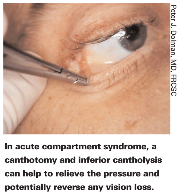
"Trauma patients need to be followed throughout the remainder of life, for two reasons," says Dr. Recchia. "First, these patients are at increased risk for ocular complications, including angle-recession glaucoma, cataract and a peripheral retinal tear as a result of their injuries. The long-term prognosis following rupture of the globe depends in part on the location of the injury. If the injury is restricted to the anterior segment but not in the visual axis, the patient may retain good vision; he may have a little astigmatism, but otherwise should do well. If the corneal wound goes through the visual axis and it's severe, the patient may eventually need a corneal transplant in order to get central vision back. If it's a scleral wound, there's a high risk of retinal detachment because of vitreous loss and incarceration causing traction on the retina. Patients in the latter situation should be evaluated promptly by a vitreoretinal surgeon once the immediate crisis is managed.
"Second, statistics indicate that patients who have trauma in one eye are likely to have trauma in the other eye and are more likely to die from trauma later in life," he says. "Clearly, some of this risk is lifestyle related. And of course, loss of one eye leads to loss of depth perception and other visual issues that could contribute to the likelihood of further accidents."
Dr. Hamill's experience supports the conclusion that trauma patients are at greater-than-average risk for further injury. "At our county hospital we went back and reviewed the risk factors for open-globe injuries," he says. "One of the highest risk factors was having had a previous open-globe injury! It seems that these are high-risk individuals indulging in high-risk behavior."
Making the Best of It
"Many ophthalmologists may not deal with trauma often enough to feel totally comfortable managing it," observes Dr. Recchia. "Everyone will have to resort to doing some research in certain cases. The key is to remember the general principles we've been discussing: Make sure there are no associated injuries; make sure the patient is otherwise stable; check whether the globe is open or closed; protect the eye from pressure; get a CT scan to look for any orbital injuries or intraorbital foreign bodies; and then treat each injury accordingly."
Dr. Dolman adds that it's important to be on the lookout for compartment syndrome. "Compartment syndrome can rapidly lead to loss of vision if not managed properly," he says. "Also, remember to check new patients' histories for past trauma. Late complications from earlier trauma are always a possibility. Last but not least, remember to advise any trauma patient to take extra steps to protect the good eye if one eye has been injured."


