A 13-year-old Caucasian female presented with complaints of a new bump in her right lower lid for seven to 10 days. She denied vision changes, pain, history of trauma or recent illness, but had begun to experience diplopia in the 24 hours preceding her presentation. Systemic review of symptoms was negative for joint pains, shortness of breath, cough, abnormal bowel movements or urinary symptoms. She had first presented to her ophthalmologist one week prior, and treatment was initiated with warm compresses and cephalexin for presumed dacryocystitis. At that time she had had a questionable right hypertropia with a 5 x 10-mm fluctuant mass beneath her nasal right lower lid; her exam was otherwise unremarkable.
Medical History
The patient had a past medical history of scoliosis and ocular history of refractive error and soft contact lens use. She denied tobacco, alcohol and intravenous drug abuse. She was not on any medications and denied any known drug allergies.
Examination
At the time of presentation she was afebrile with stable vital signs. Her external examination demonstrated a firm, non-tender, 20-mm nodule nasally beneath the right lower lid. Hertel exophthalmometry revealed 6 mm of right-sided proptosis (See Figure 1). On ocular examination, best corrected visual acuity was 20/40 OD and 20/25 OS. Pupillary exam showed no anisocoria or relative afferent pupillary defect. A 10 prism diopter right hypertropia was present in primary gaze, but extraocular motility was full bilaterally. Visual fields were full to confrontation, and Ishihara color plates were 10/10 in both eyes. Anterior slit-lamp examination and fundoscopic examination were normal; intraocular pressures were 16 mmHg OU by Goldmann tonometry.
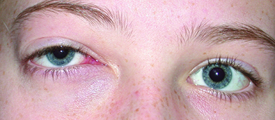 |
| Figure 1. External photography demonstrating right orbital mass lesion beneath right lower lid. |
What is your differential diagnosis? What further workup would you pursue?
Please click this link for diagnosis, workup, treatment and discussion.
Please click this link for diagnosis, workup, treatment and discussion.


