Sometimes, the best place to hide is in plain sight. This is the case with dry-eye syndrome's elusive negative effects on vision. Patients can sense that the decrease in vision is there—that something's happening to their sight—but, in many cases, the decreased vision is literally gone in a blink. So, dry-eye sufferers often don't focus on their vision, and instead present at your office with complaints of grittiness and burning.
Their physician may believe them when they say things are sometimes blurry, but, when he tries to measure the vision decrease, the patients' acuity is often confoundingly normal. In the past five to eight years or so, though, researchers have begun to develop ways to pin down the visual fluctuations associated with dry eye, and have gained new insights into the disease in the process. Here's a look at what we know.
An Elusive Problem
Michael Lemp, MD, clinical professor of ophthalmology at
"For many years, we didn't think dry eye impacted vision," he says. "This is because the vast majority of patients who would come to see us didn't complain of vision problems.
And, when we checked their vision on our conventional charts, they could read well. So, we classified dry eye as a very uncomfortable disease that can be debilitating due to its pain and discomfort, but we didn't count vision as one of the things that it threatened.
However, we knew that wasn't entirely true, because the small number of patients with more severe disease in whom the central cornea became quite irregular did have a reduction in vision."
As clinicians and researchers studied dry eye, they began to connect the interaction of the tear film and the blink response, and the patients' blurry vision became more understandable and measurable.
"We knew that, in between blinks in the normal person, you have a metastable tear film," says Dr. Lemp. "This means that the tear film is continuous, and provides a smooth surface over the cornea in between the blinks. We found that, in the interval between blinks in dry-eye patients—the inter-blink interval—they didn't maintain a continuous tear film. The average inter-blink interval is around seven seconds, but in these dry-eye patients their unstable tear film would break up at around two or three seconds. They compensated for this by, naturally, blinking more often, which made it difficult to measure acuity loss in the exam room because they can momentarily clear their vision with a blink and then read the chart with 20/20 vision. We didn't have any way of checking what their vision was at five seconds after a blink."
Researchers would soon begin to attack this very problem.
Blinking and Vision
As researchers began looking at the tear film in dry-eye patients, and its interaction with the blink, they began to get an idea of just how bad a dry-eye patient's vision can get.
"The cornea is the most powerful optical surface of the eye," remarks Mark Abelson, MD, associate clinical professor of ophthalmology at
Instead, it's sort of bumpy and granular, not the highly polished surface you'd expect in, say, an expensive camera. What makes it that highly polished surface—and sometimes even more—is that it has a fluid surface over it composed of oil on water, held by mucin, held by microvilli on the epithelial surface and continually smoothed over by the motion of the lids. If you look at the tear film immediately after a blink, it's perfection itself. This isn't so for most people immediately before the next blink, however, which occurs about eight to 12 seconds later. At that point, there exists all kinds of diffraction on the surface."
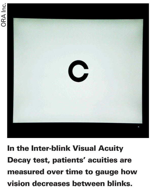
In an effort to measure dry-eye patients' vision in real time, Dr. Abelson, Pamela Walker, MD, George Ousler and their co-workers had dry-eye patients stare at a screen on which a series of Landolt Cs symbols were presented every second or two. Using a keypad, which enables a reaction time that's quicker than a verbal response from the patient, the patient indicated which direction the opening of the C shape was pointing. The system continues to increase the size of the Cs, representing different levels of visual acuity.
This process gave the researchers an idea of patients' acuities over the inter-blink interval, and they called the system the IVAD, short for Inter-blink Interval Visual Acuity Decay.
"Let's say the patient doesn't blink for eight seconds," says Dr. Abelson. "I can run through four or five different visual acuities over that eight-second time span and track their consistency. It may be 20/20 all the way through, which would be a completely normal eye. However, it might decay every second or two from 20/20 down to 20/60 or worse … So, if I'm 20/20, I'm not 20/20 for all 12 seconds that I keep my eye open if I have dry eye. I'm 20/20 for maybe four seconds, then 20/40 for another second, 20/50 for two more, 20/60 for a second or two and then maybe 20/80 before I have to blink again."
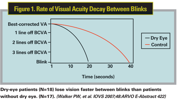
In the study using the IVAD, there were 18 dry-eye patients and 17 age-matched controls. The researchers found that dry-eye patients could only maintain their best-corrected visual acuity for 8.75 ±6.6 seconds before it started to decay, while normal patients maintained it for a statistically significant longer period of time: 19.46 ±15.97 seconds. (See Figure 1) Acuity could drop to as low as 20/80 in some patients. (Walker PW, et al. IOVS 2007;48:ARVO E-Abstract 422)
Dr. Abelson says that some dry-eye patients' vision would decrease within two seconds after blinking. "We also found a diurnal curve associated with this decay in visual acuity," he says. "So as the day progressed and the patient was fatigued, his visual function also got worse toward the end of the day. Eighty percent of dry-eye patients are worse in the evening, but everyone else is worse in the evening too. Even in normals, their corneal staining progresses throughout the day."
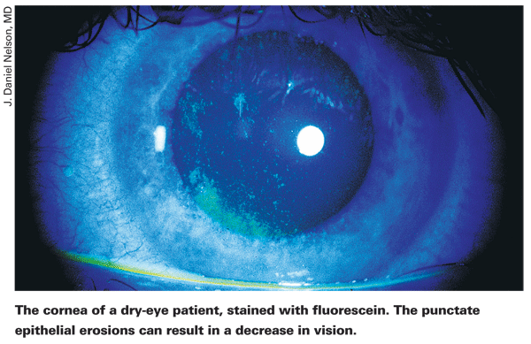
In addition to these patients with tear-film instability, there are also those patients with severe disease who actually have corneal damage. "You can have cellular damage or disruption to the surface epithelial cells," says J. Daniel Nelson, professor of ophthalmology at the
Other studies have also taken a look at dry eye's impact on vision, both quantitatively and qualitatively.
One study found a measurable adverse impact of dry eye on several activities of daily life. The study employed a questionnaire that touched on the extent to which dry-eye symptoms limited various tasks. When compared to patients without the disease, patients with dry eye were more likely to report problems with reading, working, computer use, watching television and driving (both at night and during the day).1
In another study of preop refractive surgery patients with low refractive errors, treating their dry eye actually improved their uncorrected vision. In a retrospective fashion, surgeons placed silicone punctal plugs in 21 eyes of 12 patients with dry eye. At one month, two eyes had improved by three lines, nine had improved by two, seven had improved by one and three eyes were unchanged.2
Dealing with the Problem
Though the effects of dry eye on vision are well-documented, physicians say there are still some pitfalls to avoid when dealing with a dry-eye patient who happens to complain of blurry vision.
Dr. Abelson says to be careful ascribing too much weight to a patient's blink rate.
"Clearly, the blink is the first line of defense," says Dr. Abelson. "As the disease gets more severe, however, their comfort actually increases because their sensation decreases. So, their blink pattern looks a little more normal, but then all of their signs will start to explode. They've overwhelmed their first line of defense, the blink, which apparently is driven by sensation." In one study performed by Dr. Abelson, patients who were relatively new to dry eye felt very uncomfortable but didn't have any signs of the disease. But there was another group who had the disease for almost twice as long who had had stopped experiencing symptoms and only had signs of the disease. (Casavant J, et al. IOVS 2005;46:ARVO E-Abstract 4455) "This is where you start to see the bad vision with the severe cases," says Dr. Abelson. "They have a longer inter-blink interval now, but are the last people in the world who should have one." The visual decrease will be more constant and measurable in this group.
"Also, make sure the dry-eye patient's vision is optimal," reminds Dr. Nelson.
"Sometimes these patients will have had a refraction that was done during a period when their tear film was either stable or unstable that doesn't represent how it is now.
You want to make sure you're not missing obvious optical changes. For instance, they may not have been refracted in four years. Also, they may be experiencing visual changes due to cataract. Finally, a lot of Sjögren's patients may be on Plaquenil (hydroxychloroquine). This drug can affect the macula. So, someone who's been on it for a long time may have macular changes that are causing central vision changes unrelated to dry eye."
Since most ophthalmologists won't have equipment in place to drill down into all the minute causes of decreased acuity in dry eye, Dr. Nelson says a good diagnostic treatment trial is best—treat them and see how they do. "You can put an artificial tear in the eye and see if the vision clears. Or, you could do a hard contact lens refraction to take the tear film and corneal changes out of the equation. Some physicians may have corneoscopy or corneal topography available. If so, you can use one of these methods to image the eye before and after instillation of an artificial tear to demonstrate the dry-eye changes. The key is, if they have significant dry eye and come in with visual complaints, you can treat the dry eye, increase the artificial tears, and/or add an anti-inflammatory agent and reassess the vision further down the road. If you do that, you'll probably take care of 90 percent of these patients."
1. Miljanoviæ B, Dana R, Sullivan DA, Schaumberg DA. Impact of dry-eye syndrome on vision-related quality of life. Am J Ophthalmol 2007;143:3:409-15.
2. Khalil MB, Latkany RA, Speaker MG, Yu G. Effect of punctal plugs in patients with low refractive errors considering refractive surgery. J Refract Surg 2007;23:5:467-71.