Christopher Singh, MD, Asheesh Tewari, MD, Detroit and Gaurav K. Shah, MD, St. Louis
Patient expectations following cataract surgery have been rising rapidly with technological advances in cataract and refractive surgery. Presbyopia-correcting intraocular lenses are among the latest in new technology available to patients. These have set the bar higher for surgical outcomes, with spectacle independence at near as well as at distance now being the desired outcome.
With these lenses gaining popularity with surgeons and patients alike, a new set of challenges is presented to the vitreoretinal surgeon. Performing vitreoretinal surgery through these lenses can be challenging for a variety of reasons, but with careful preoperative and intraoperative planning, these challenges can be met, maintaining the best possible patient outcomes.
Crystalens
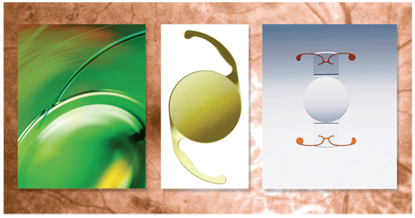
Crystalens (Eyeonics, recently acquired by Bausch & Lomb) is an accommodating monofocal lens composed of a biconvex silicone plate. When a patient accommodates, the single focal point is designed to move anteriorly on hinged haptics, which typically provides +1.25 D. When accommodation is relaxed, the optic moves posteriorly, allowing for distance vision.1 This lens functions correctly when placed in the capsular bag.
Challenges to the vitreoretinal surgeon with this lens in place include the silicone composition, intraoperative optic mobility and intraoperative condensation. This lens should not be placed in diabetics, as they may need future vitrectomy surgery with silicone oil, which would adhere to the optic. The other current presbyopia-correcting IOLs are composed of acrylic, and may be more appropriate in these cases.
The mobility of the Crystalens can present a challenge intraoperatively, particularly if the posterior capsule has previously been compromised. In order to function properly, the optic and haptics must remain in the capsular bag. The optic must also be centered for ideal function. During air-fluid exchange, the optic can vault anteriorly into the iris plane. This is a necessary step in vitreoretinal surgery and lens vaulting may not be avoidable. In our experience, by leaving the eye relatively soft following nonexpansile gas fill, the optic will return to its native position in the bag. If this step is not taken, there is risk of iris capture of the optic or a malpositioned lens. This may result in another surgical procedure to reposition the lens.
Compromised View
Condensation on the optic can happen, particularly in an eye that has had posterior capsulotomy. This, combined with a mobile Crystalens optic, can severely compromise the view following air-fluid exchange. Coating the posterior aspect of the IOL with a viscoelastic can diminish condensation. Additionally, placing the air infusion line in ice or using a humidifier may help prevent this problem. If condensation does occur, viscoelastic can be used to improve the view, but it is more advisable to take preventive steps.
Multifocal IOLs
The multifocal IOLs include ReSTOR (Alcon) and ReZoom (Advanced Medical Optics). These are acrylic 3-piece IOLs with concentric circular zones. The multiple circular zones provide two lens powers to the patient, distance and near. When looking in the distance, the near images are defocused and the patient sees only the distance images. When a patient looks at a near object, approximately +3.5 D are added from the near zones and the distance images are defocused. By using characteristics of both lens powers, patients are also able to achieve satisfactory intermediate vision.2,3
The concentric optical zones represent a noteworthy advance in spectacle independent vision, but present the vitreoretinal surgeon with a significant challenge. Macular holes, epiretinal membranes and diabetic retinopathy typically require delicate membrane peeling at the surface of the macula. During these maneuvers, there is risk of iatrogenic trauma to the surface of the macula.
Surgical Issues
Through a multifocal IOL, there are major changes in depth perception when moving between the optical zones. This increases the risk for iatrogenic macular trauma and necessitates alternative strategies. When peeling macular membranes through a multifocal IOL, surgeons are strongly encouraged to stay within one zone. Start the membrane peel by using a pinch and grab technique in the parafoveal area. Peel the membrane to the edge of the central circular zone only. If further peeling is required, start in the next most peripheral zone, and peel a strip circumferentially within that zone only. Avoiding the prismatic effect is the safest approach, in our experience.
Performing vitreoretinal surgery through presbyopia-correcting IOLs presents the surgeon with a new set of challenges. It is necessary for the vitreoretinal surgeon to have a new skill set to meet these challenges. As these IOLs gain popularity and become more commonplace, adaptive surgical techniques for posterior work must become commonplace as well to provide patients with the best possible outcomes.
Dr. Singh is a vitreoretinal fellow at the Kresge Eye Institute, Wayne State University Department of Ophthalmology. Dr. Tewari is a retina specialist at the Kresge Eye Institute and is an assistant professor with the Wayne State University Department of Ophthalmology. Dr. Shah is from the Barnes Retina Institute and is a clinical associate professor with Washington University Department of Ophthalmology and Visual Science. Contact Dr. Tewari at Kresge Eye Institute, 4717 St. Antoine,
1. Macsai MS, Padnick-Silver L, Fontes BM. Visual outcomes after accommodating intraocular lens implantation. J Cataract Refract Surg. 2006;32:628-633.
2. Alfonso JF, Fernández-Vega L, Baamonde MB, Montés-Micó R. Prospective visual evaluation of apodized diffractive intraocular lenses. J Cataract Refract Surg. 2007;33:1235-1243.
3. Chiam PJ, Chan JH, Haider SI, et al. Fucntional vision with bilateral ReZoom and ReSTOR intraocular lenses 6 months after cataract surgery. J Cataract Refract Surg. 2007;33:2057-61.


