COVID-19 is changing the role of portable retinal imaging devices. With restricted clinic options, telemedicine, and the need to maintain social distance and reduce both staff and patient volume, handheld devices have found an expanding niche. “We’ve had to deploy services in spaces without standard imaging devices due to space limitations,” says Delia Carbrera DeBuc, PhD, a research associate professor of ophthalmology at Bascom Palmer in Miami. “Portable devices are penetrating the markets now. We’re also seeing an increase in demand for virtual and hybrid visits at Bascom Palmer.”
Here, Dr. Cabrera DeBuc shares some insight on portable retinal cameras in today’s clinic, and we’ll review 10 options for visualizing the retina in atypical clinic settings.
Handheld Retinal Cameras
Handheld retinal cameras have a number of applications, from community screening and triage to pediatric and geriatric care, where patients may not be able to sit comfortably at a slit lamp or traditional fundus camera. The image quality and field of view of a portable device are variable and generally held to be inferior to those of standard fundus cameras, but some studies have shown that portable fundus cameras can yield professional-quality fundus images.1
Dr. Cabrera DeBuc says the main issue with handheld cameras is stability. “Getting a good image can be difficult,” she says. “It’s best if you have a trained technician. Many of the portable cameras I’ve used have slit lamp attachments, which I prefer when I use them.”
Additionally, handheld cameras provide a smaller field of view, so you may have to take multiple photos, and some details may be skewed. “You can’t see all the details like you can on a standard fundus machine,” she explains. “Portable cameras are good for a first indication to have an idea of whether or not you should refer the patient for better analysis.”
If you need a handheld retinal camera during COVID-19, Dr. Cabrera DeBuc says that sanitation and safety are key. “The main concern is contact,” she says. “We haven’t yet developed an at-home device for retinal selfie imaging, like the at-home tonometers. The technician needs to face the patient, so you should use a shield if possible.”
Though portable retinal cameras are smaller than traditional fundus cameras, they require more direct contact for imaging. “Most portable cameras have an eye cup, which comes in direct contact with the patient,” she says. “Other fundus machines have a lens that you just move forward and back to focus, but the eye cup sits against the patient’s eye the entire time to ensure the correct positioning. From a sanitation standpoint, I think the handheld cameras require more careful cleaning.”
Dr. Cabrera DeBuc says that if you’re thinking of incorporating a portable retinal camera in your clinic to accommodate COVID-19 restrictions or for teleophthalmology, becoming comfortable with the camera or having training is key. “The main complaint with handhelds is that we’re not able to get good images,” she says. “Some people are better with the cameras than others, though. Your ophthalmic technician may be the right person to get properly trained on a handheld camera. It’s also a good idea to ask the product developer for assistance. They know a lot of tips and tricks for using the camera that you may not be familiar with.”
Here’s a brief overview of five portable fundus cameras.
• Dragonfly (Eyefficient; Aurora, Ohio) is a lightweight, handheld fundus camera. Its nonmydriatic technology eliminates 20 minutes of patient dilation time, says the company. The Dragonfly offers a 45-degree field of view for checking critical areas of the retina and includes a 3.97-inch touch screen and five internal fixation targets for capturing retinal images. With built-in Wi-Fi, micro SD and Miracast connectivity, the Dragonfly offers quick and easy dissemination of retinal images through the Web from remote locations, says Eyefficient. A rechargeable lithium-ion battery is included. For information, visit eyefficient.com.
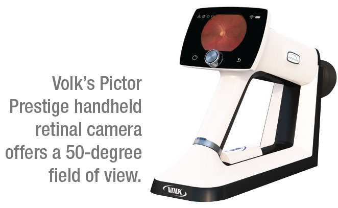 |
• Pictor Prestige (Volk) is a portable retinal camera specifically designed for imaging small pupils—as small as 3 mm—with a 50-degree field of view. Volk says the device’s image quality analysis ensures that you won’t need a repeat imaging visit. The Pictor Prestige features a user-friendly interface, a battery lasting a full day in the clinic and a backup battery for overtime use. For information, visit volk.com.
• VersaCam α (Nidek) is a portable fundus imaging unit with autofocus and autoshot options for button-free use, in addition to a manual capture mode. The VersaCam weighs 445 g (about 1 lb.) and comes with a rechargeable battery that holds a charge for about three hours of continuous operation, according to Nidek. The camera itself is a 5-megapixel clear-color camera with 45-degree horizontal by 40-degree angles of view and seven internal fixation lamps for stable fixation during measurement. Data and image review appears on a 3.5-inch touch screen, and data can be saved on an SD card or transferred to image software for Nidek devices. There’s also an optional slit lamp attachment. For information, visit nidek.intl.com.
• Signal (Topcon) is a lightweight, compact retinal camera, making it ideal for visiting bedridden patients or those in their own homes, the company says. Signal enables ophthalmologists to perform non-mydriatic retinal examinations with a 50 x 40-degree field of view. The camera produces true-color fundus images and features nine fixation targets for central and peripheral imaging. The autofocus function features speed image acquisition, says Topcon. The device can operate for approximately five continuous hours and images can be uploaded to the cloud or stored in the device’s memory. In addition to being mobile, Topcon says Signal has low brightness and intensity that can help when working with small children, who are often scared by the bright lights of stationary retinal cameras. Optional slit lamp adapters are available. For information, visit topconmedical.com.
• VisuScout 100 (Zeiss) features a 5-megapixel handheld fundus camera that takes color and red-free images with a 40-degree field of view and meets all relevant ISO 10940 fundus camera standard requirements. The VisuScout’s nonmydriatic operation (minimum pupil size 3.5 mm) and autofocus capabilities make dilation unnecessary, says the company. Nine internal fixation LEDs facilitate patient alignment and the capture of peripheral images. Zeiss says the VisuScout is easy to operate, requiring only a brief training session, and that examinations can be performed quickly. The device comes with a rechargeable lithium-ion battery. Data and images can be uploaded with Wi-Fi or by USB, or saved on an SD card. For information, visit zeiss.com.
Smartphone Retinal Imaging
Smartphone ophthalmic imaging has become increasingly popular, especially in less-advantaged regions, due to phones’ portability, ubiquity, low cost and diagnostic quality that’s comparable to that of traditional fundus camera images.2 “Many places around the world don’t have enough specialists or resources, so these tools are helpful,” Dr. Cabrera DeBuc says. “If they can use a lens adapter with a 20-degree field of view to see the back of the eye, they can at least have an idea of whether or not the patient needs to be referred.”
Smartphone retinal imaging’s potential in teleophthalmology is also growing, with integrated smartphone apps that allow image and file sharing paired to EHRs. Here are a few adapter options for your smartphone.
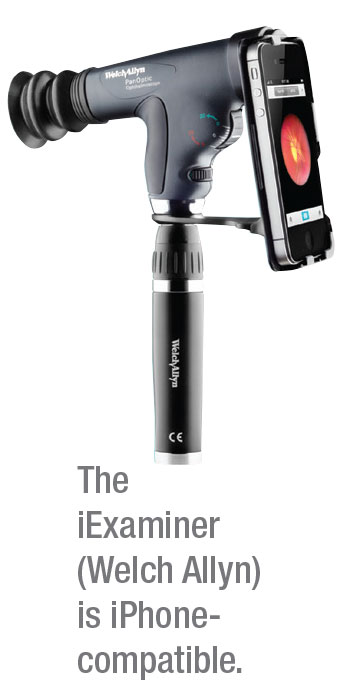 |
• iExaminer (Welch Allyn; Skaneateles Falls, New York) is a non-mydriatic fundus imaging device for iPhone 6, 6s and 6-plus models. The iExaminer incorporates the company’s PanOptic ophthalmoscope for capturing high-resolution images with a view of the fundus five-times larger than the standard Welch Allyn ophthalmoscope, and a 25-degree field of view of the fundus. An accompanying app enables file storage, sharing via email and printing. Free and pro versions of the app are available in the App Store. For information, visit welchallyn.com.
• Peek Retina (Peek Vision, London) is a small, three-part modular adapter for Android-based devices that can be applied to a phone in less than 30 seconds, according to the company. With Peek, doctors can immediately share files, transfer them to EHRs or show patients what they’ve seen for an educational experience.
The Peek Retina device underwent a validation trial of 2,152 optic nerve images in 2016. The researchers found that nonclinical photographers were able to use the low-cost smartphone adapter to acquire images sufficient to enable independent remote grading comparable to that done using images acquired by an ophthalmic assistant using a desktop retinal camera. For information, visit peekvision.org.
• D-Eye (D-Eye; Padova, Italy) is a digital direct ophthalmoscope that attaches to a smartphone (iPhone 5s, SE, 6-generations, 7 or 8 ) and takes high-quality photos and video of the fundus. D-Eye doesn’t require dilation, and offers a 6-degree field of view with undilated pupils for viewing the optic nerve and posterior pole. With dilated pupils, users can achieve a 20-degree field of view. The adapter fits over a smartphone’s camera aperture and uses the phone’s LED light. A companion D-Eye app allows doctors to enter patient information, focus the retinal camera and record, archive, view and transmit images.
The company says that D-Eye is ideal for telemedicine diagnoses and consultations. The FDA-approved device also uses a HIPPA-compliant cloud-based data management platform to store and share images for evaluation and screening. For information, visit d-eyecare.com.
• Paxos Scope (DigiSight Technologies; San Francisco) is an FDA-registered portable ophthalmic camera that enables image capture of the anterior segment and retina. Paired with the Paxos mobile app, ophthalmologists can coordinate with others in real-time by using the team collaboration features and also perform documentation in EHRs. The Paxos platform enables provider-to-provider teleconsultations for eye-related disease, says the company.
• Fundus on Phone (Remidio; Bangalore, India) is a nonmydriatic, smartphone-enabled retinal camera with integrated artificial intelligence. Its optics design is FDA-approved, and the device comes with a user-friendly app for accessing patient data and EHRs. This fundus camera also includes a diabetic retinopathy screening solution with an offline AI for DR detection within 10 seconds, according to the company. One study concluded that the FOP can be an initial tool for mass retinal screening for those with diabetes.3 For information, visit remidio.us/fop.php. REVIEW
Dr. Cabrera DeBuc has no financial interests in any of the products mentioned.
1. Jin K, Lu H, Su A, et al. Telemedicine screening of retinal diseases with a handheld portable non-mydriatic fundus camera. BMC Ophthalmol 2017;17:1:89.
2. Adam MK, Brady CJ, Flowers AM, et al. Quality and diagnostic utility of mydriatic smartphone photography: The smartphone ophthalmoscopy reliability trial. Ophthalmic Surg Lasers Imaging Retina 2015;46:631-637.
3. Rajalakshmi R, Subashini R, Mohan Anjana R, et al. Automated diabetic retinopathy detection in smartphone-based fundus photography using artificial intelligence. Eye (Lond) 2018;32:6:1138-1144. [Epub ahead of print].