Here, you’ll find a sample of some of the more recent options designed to help surgeons achieve accurate toric alignment.
IntelliAxis-L System
One of the newest entries in this area is the IntelliAxis-L system, now part of the latest upgrade to the Lensar Laser System (Lensar), which was showcased at the American Academy of Ophthalmology meeting in November 2017. The company describes the laser as the only one on the market
 |
| The IntelliAxis-L System is a new upgrade now available for the Lensar Laser System. |
IntelliAxis-L uses Lensar’s diagnostic capabilities, iris registration software and intraoperative imaging to precisely and “permanently” identify the location of the steep corneal axis at the capsular plane for toric IOL alignment. The system creates custom marks, which the company says can’t be duplicated by any other device currently available, which remain visible postoperatively to help identify any unexpected rotation and help guide any needed realignment of the toric IOL to its optimal position.
The company notes that the Lensar System’s astigmatism-management features include:
• integration of all corneal measurements, including total corneal refractive power and total corneal astigmatism, to guide placement of arcuate incisions;
• iris registration with automatic cyclorotation adjustment;
• arcuate incision planning; and
• surgeon tables for managing pre-existing and surgically-induced astigmatism.
For more information, visit: lensar.com/features.php
Tocular System
The Tocular System (Haag-Streit Surgical) is a rotatable, wide-angle ocular with a clearly visible integrated reticle in the center of the visual field. The device can be rotated manually via a sterilizable knob, and it can achieve IOL alignment within two degrees of the target axis. The eye must be marked preoperatively using a marking system of your choice. (Haag-Streit recommends the Davis premarking system, available from John Weiss.) After the IOL is implanted, the surgeon adjusts the microscope zoom to visualize the ink marks, and then rotates the Tocular reticle to align with them. The IOL can then be aligned using the medium and small reticle markings as a guide.
For more information visit: haag-streit.com/fileadmin/Haag-Streit_Surgical/Download_documents/Brochures/Flyer_TOCULAR.pdf
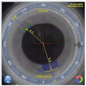
Verion Image Guided System
The Verion’s Reference Unit (Alcon) is a modified keratometer that takes corneal power measurements, captures a high-resolution reference image, automatically detects scleral vessels, as well as the limbus, the pupil and iris features, and then shows where the steep axis is relative to a picture of the eye. The data c
 |
| The Verion Image Guided System is compatible with most surgical microscopes. |
The Digital Marker software tracks the eye, providing a digital overlay to help with alignment. It can also provide guides for relaxing incisions, creating the capsulorhexis and positioning a multifocal lens. For more information, visit: myalcon.com/products/surgical/verion-guided-system/
RoboMarker System
According to the manufacturer (Surgilum), the RoboMarker System is the first self-leveling corneal marker with pre-inked, sterile, disposable tips and an integrated fixation light. The device has a concealed dual-pendular weight system balanced between two high-precision ball-bearings that ensures that the RoboMarker ring dial will maintain your chosen axis to within one degree.
For marking the cornea, the system provides single-use, sterile “RoboTips”— “fins” that come pre-applied with a dry marking formula. When the dry formula contacts the tear film, it produces precise marks that last up to two hours, long enough for preoperative marks to still be clear in the
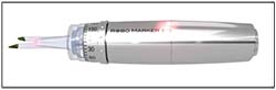 |
| The RoboMarker System from Surgilum. |
The company claims that this marking system can save time compared to some multistep marking protocols. For more information visit: surgilum.com/products/robomarker/
EyeSuite Toric Planner
The EyeSuite Toric Planner (Haag-Streit) enables the surgeon to intuitively plan the surgery using high-resolution images of the eye. It also features an incision-optimization tool to help the surgeon minimize residual astigmatism by placing the incision in the right spot. The Planner is part of Haag-Streit’s T-Cone Toric Platform, an optional addition to the LenStar biometer. The T-Cone Toric Platform complements the LenStar’s comprehensive measurement spectrum with 11-ring placido topography, to help confirm the axis location and the regularity and symmetry of the astigmatism. Toric IOL calculation is accomplished with the integrated Barrett Toric Calculator.
Planning the operation using high-resolution eye images allows the user to define additional guiding lines to anatomical landmarks that will be recognizable in the intraoperative view. The planning sketch can be printed and hung near the microscope.
For more information, visit: haag-streit.com/haag-streit-diagnostics/products/biometry/t-cone-toric-platform/
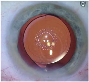
Cataract Suite Markerless
The Cataract Suite Markerless from Carl Zeiss Meditec is a set of interconnected instruments that enables—among other things—toric IOL and limbal relaxing incision alignment without the need to mark the eye. Data transfer between instruments is managed by Zeiss’s FORUM data-management system, eliminating the need for manual transfer of information and greatly increasing surgical efficiency, according to the company.
After the IOLMaster takes its
 |
| Zeiss’ Cataract Suite Markerless works with the IOLMaster and Callisto eye system. |
This function is integrated into Zeiss’s OPMI Lumera 700 microscope; for all other current Zeiss surgical microscopes, it’s available as a retrofit. For more information, visit: zeiss.com/meditec/us/products/ophthalmology-optometry/cataract/zeiss-cataract-suite-markerless.html
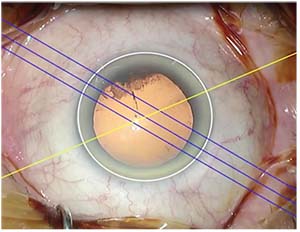
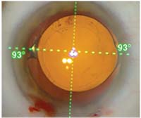
Illuminating Surgical Keratoscope V5
The fifth-generation Illuminating Surgical Keratoscope (Mastel) features visual axis fixation/centration and lights indicating the primary meridian, adjustable from 0 to 180 degrees. The device is designed to ac-count for cyclotorsion. The surgeon marks the eye at 3, 6 and 9 o’clock with the patient upright. Intraoperatively, while the patient fixates on a pulsating red light, the surgeon aligns the LED cardinal references with the pre-marks and sets the primary meridian to the desired position. The device can be attached to most surgical microscopes, with standard sizes for Zeiss and Leica scopes.
The manufacturer notes that this device is not intended as a sole indicator of absolute alignment, but as a complement to a s
 |
| When using the Illuminating Surgical Keratoscope V5, the two reflections reveal the toricity of the lens and cornea, helping the surgeon to align the lens. |
Discovery System
The Discovery System (Innovative Visual Systems) measures multiple eye characteristics including white-to-white, pupil diameter, corneal topography and both corneal and whole-eye higher-order aberrations, using wavefront technology. The system provides toric IOL axis orientation (to ANSI precision standards), while iris registration corrects for head tilt and/or cyclorotation. Together, retroillumination photography and iris registration enable precise, direct assessment of IOL orientation. The Discovery System also provides postoperative astigmatism analysis and repositioning guidance, showing how to correct any error in toric IOL alignment. In addition, once the surgeon has entered the patient’s corrected axis, the Discovery System will display a simulated image equivalent to a 20/30 letter on the Snellen chart, showing the expected visual acuity after IOL repositioning.
For more information, visit: ivisualsystems.com/index.html
Osher Toric Alignment System
This system (from Eye Photo Systems) employs iris fingerprinting to improve the accuracy of toric implant alignment. A detailed, high-resolution photograph of the dilated pupil is captured during the initial preop examination. The keratometric meridians and the IOL goal line, transposed from the toric IOL calculator, can be overlaid on the image in the form of a protractor. That image can then be brought into the OR on a USB drive or printed as a hard-copy photograph for reference during the IOL alignment. The surgeon locates at least two unique landmarks in the iris such as crypts, nevi or brushfields. During surgery, those landmarks act as guides to help the surgeon use a Mendez guiding ring to accurately match the target meridian shown in the image.
For more information, visit: eyephotosystems.com/toric-alignment/
Wet-Field Osher ThermoDot Marker
The Osher ThermoDot Marker (Beaver-Visitec International) is a bipolar intraoperative instrument that makes a tiny pinpoint cautery mark on the cornea, eliminating the need for ink. The fine dot(s) can be used as a precise reference point for a variety of procedures, i
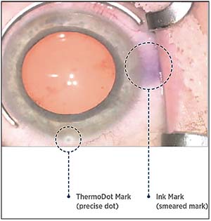 |
| The Osher ThermoDot device creates a tiny, painless cautery mark that will not smear, but disappears after a day or two. |
For more information visit: beaver-visitec.com/upload/BVI_SellSheet_WetField_Osher_ThermoDot_LowRes.pdf
Optiwave Refractive Analysis System, with VerifEye+
The ORA intraoperative system (Alcon) measures the eye’s refractive state while the patient is on the table, providing continuous assessment and recommendations for lens power—including the axis and magnitude of astigmatism. Because this approach measures the total refractive state of the eye, including posterior corneal astigmatism, it helps to improve astigmatic outcomes, the company states.
The system provides overlays showing the optimum positioning for limbal relaxing incisions, and then provides continuous validation of LRI
 |
| The ORA intraoperative system shows real-time streaming data, taking posterior corneal astigmatism into account. |
TrueGuide
TrueGuide (TrueVision 3D Surgical) is a software application that generates templates in the surgeon’s surgical field of view, in real time, to aid in IOL alignment and incision creation, employing customized surgeon profiles. It uses autoregistration with a preoperative image and 3-D eye-tracking that helps to eliminate parallax errors during the alignment.
TrueGuide provides real-time vector analysis for incision optimization and toric IOL and LRI positioning, taking all variables into account; it
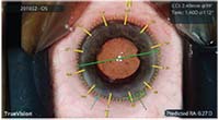 |
| The TrueGuide system generates templates in the surgical field of view in real time. |
Visit truevisionsys.com/trueguide.html for more information. REVIEW


