A 55-year-old man with a history of soft contact lens wear presented to the Wills Eye Cornea Service for evaluation of six weeks of pain and blurry vision in the right eye. The patient initially presented to an outside optometrist who diagnosed a corneal abrasion in the right eye. Two weeks later he presented to an outside ophthalmologist with worsening symptoms in his right eye, was diagnosed with a corneal ulcer, and was initiated on moxifloxacin 0.5% four times per day. He continued to worsen, and three days later he was started on fortified vancomycin drops every hour while awake, oral valacyclovir 500 mg three times per day, and topical trifluridine nine times per day, and corneal cultures were obtained. Cultures were negative for bacteria and fungi. Three weeks later the patient presented to the Wills Eye Cornea Service due to persistence of his symptoms. Prior to presenting to Wills, the patient had at one time been on the following treatments: loteprednol 0.5%; moxifloxacin 0.5%; fortified vancomycin; trifluridine; tobramycin/dexamethasone; valacyclovir; doxycycline; and artificial tears.
Medical History
At his initial visit to Wills Eye, the patient denied contact lens overwear, sleeping in lenses, showering or swimming in lenses. His ocular history was otherwise negative. His past medical history was significant only for hyperlipidemia. His only systemic medication was a statin.
Examination
The patient’s visual acuity with correction was 20/100 in the right eye and 20/20 in the left eye. His pupils were equal and reactive with no afferent pupillary defect. His motility was full, and intraocular pressures were 8 mmHg bilaterally.
What is your differential diagnosis? What further workup would you pursue?
- See more at: http://www.revophth.com/content/d/wills_eye_resident_case_series/i/2548/c/42794/#sthash.1oQnKdhN.dpufWhat is your differential diagnosis? What further workup would you pursue?
- See more at: http://www.revophth.com/content/d/wills_eye_resident_case_series/i/2548/c/42794/#sthash.tKH5GvEM.dpufWhat is your differential diagnosis? What further workup would you pursue?
- See more at: http://www.revophth.com/content/d/wills_eye_resident_case_series/i/2548/c/42794/#sthash.tKH5GvEM.dpuf
|
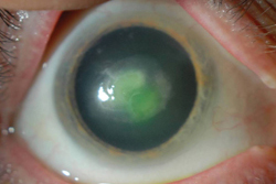
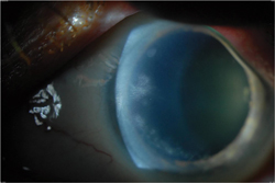
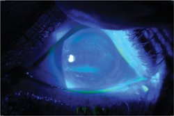
On slit-lamp examination, the right eye revealed 1+ injection and a 3.75 mm x 4 mm infiltrate with an overlying epithelial defect (See Figure 1). The left eye had trace injection, scattered peripheral subepithelial corneal infiltrates, and a pseudo-dendritic epithelial staining pattern (See Figures 2 & 3).
What is your differential diagnosis? What further workup would you pursue?
Please click this link for diagnosis, workup, treatment and discussion.
Please click this link for diagnosis, workup, treatment and discussion.
What is your differential diagnosis? What further workup would you pursue?
- See more at: http://www.revophth.com/content/d/wills_eye_resident_case_series/i/2548/c/42794/#sthash.1oQnKdhN.dpufWhat is your differential diagnosis? What further workup would you pursue?
- See more at: http://www.revophth.com/content/d/wills_eye_resident_case_series/i/2548/c/42794/#sthash.tKH5GvEM.dpufWhat is your differential diagnosis? What further workup would you pursue?
- See more at: http://www.revophth.com/content/d/wills_eye_resident_case_series/i/2548/c/42794/#sthash.1oQnKdhN.dpufWhat is your differential diagnosis? What further workup would you pursue?


