Presentation
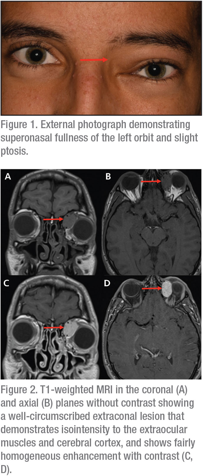 |
An 18-year-old Caucasian male presented with a painless nodule in the superior medial canthus (See Figure 1). The patient denied vision changes, diplopia or rapid growth of the lesion. The patient first noticed the mass 11 months prior and initially saw his primary medical doctor, who referred the patient to an ophthalmologist. Four months later, on ophthalmic examination, he was diagnosed with allergic conjunctivitis. Allergy eye drops and warm compresses were prescribed twice daily for a month with no improvement. On follow-up, orbital imaging was recommended. Computed tomography revealed a well-circumscribed, extraconal mass within the superonasal left orbit, measuring 24 mm x 12 mm x 17 mm and without involvement of the extraocular muscles, but with mass effect on the medial rectus muscle and globe. The CT attenuation of approximately 60 Hounsfield units suggested a solid or complex cystic mass. Magnetic resonance imaging demonstrated intermediate T2 and T1 signal and fairly homogeneous contrast enhancement with no cystic spaces or fluid levels within the mass (See Figure 2). The patient was referred to the Wills Eye Hospital Ocular Oncology Department for further management.
Medical History
There was no relevant past medical, ocular history or prior surgery. There was no trauma to either eye. Family history was noncontributory.
Examination
Ocular examination demonstrated visual acuity of 20/30 (PH 20/20-1) in the right eye and 20/30 (PH 20/20) in the left eye. Pupils were equal, round and reactive bilaterally with no afferent pupillary defect. Intraocular pressures were 10 and 13 mmHg OD and OS, respectively. Color plates were full in both eyes. Extraocular motility was full OU. Visual fields were full to confrontation OU. External examination showed a non-tender, non-erythematous palpable mass measuring approximately 20 mm x 15 mm at the left medial orbital rim, with displacement of the globe inferotemporally and 2 mm ptosis of the left upper eyelid. Hertel exophthalmometry showed no proptosis. Anterior segment and funduscopic examination were normal OU.
What is your diagnosis? What further workup would you pursue? Please click this link for diagnosis, workup, treatment and discussion.


