Cornea specialists continue to perfect their technique and improve their outcomes with endothelial keratoplasty procedures. Along with heightened interest in the role of anti-VEGF drugs, these are the key areas of focus in the year's ARVO report.
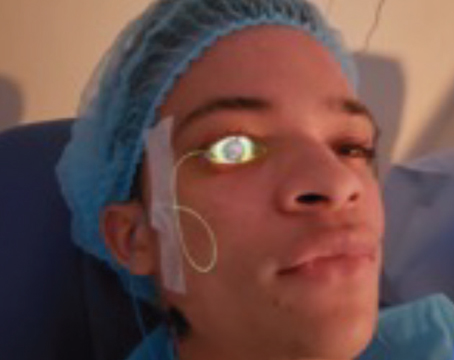
DSEK
Descemet's stripping endothelial keratoplasty provides rapid visual rehabilitation and excellent BCVA in patients with endothelial dysfunction, but requires multiple postop examinations immediately after DSEK in order to diagnose and treat complications, say a pair of researchers at the
Fifty-four eyes underwent DSEK only, 37 underwent combined phacoemulsification and DSEK, three underwent DSEK and anterior vitrectomy, one underwent DSEK and synechiolysis, one underwent DSEK and superficial keratectomy, and one underwent DSEK and Ahmed shunt revision. The incidence of postop pupillary block was 20 percent. One patient developed aqueous misdirection requiring pars plana vitrectomy and peripheral iridectomy. The incidence of lenticule dislocation was 25 percent, all requiring lenticule repositioning and air tamponade. The incidence of iatrogenic graft failure was 3 percent and allograft rejection, 1 percent, all requiring repeat DSEK (See Table 1.)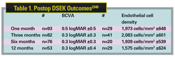
Despite leaving just enough air in the anterior segment to cover the disc edges, there was a high rate of postoperative pupillary block and lenticule dislocation in this series. 2340
A team from the
Preliminary results of a small series of cases at the
Higher-order anterior corneal aberrations between three keratoplasty techniques (conventional PK, four eyes/four patients; femtosecond laser with a "zig-zag" pattern incision, five eyes/five patients; and DSEK, four eyes/four patients) were compared using Zernike polynomial values for 3rd- to 8th-order aberrations obtained from Pentacam corneal topography. Both conventional PK and femtosecond techniques were performed with a 24-bite running suture.
Coma values were 2.2042 with the femtosecond laser "zig-zag" incision, 0.9608 with DSEK, and 3.5254 with conventional PK six months to two years postop. There was no statistically significant difference when comparing spherical aberrations or other higher (4th to 8th order) aberrations between the three techniques. 2320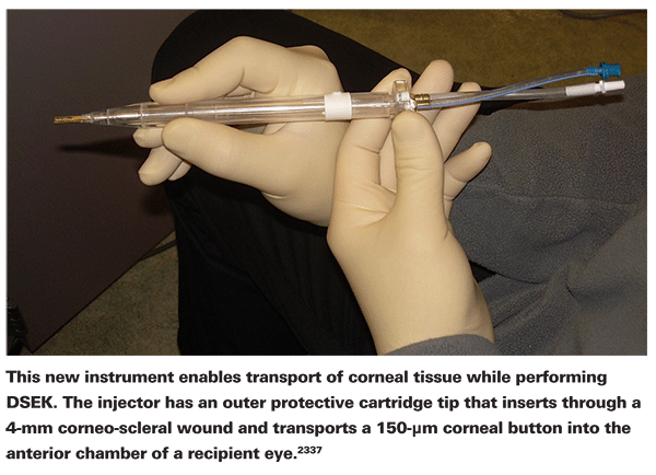
Backscattered light from the cornea decreases after DSEK, yet remains higher than normal in the posterior two-thirds of the cornea, the region that includes the interface, through six months, says a group at the Mayo Clinic College of Medicine,
In this prospective, non-randomized study, 21 eyes of 19 participants, all with corneal edema from Fuchs' dystrophy, were evaluated before surgery and at one (n=19), three (n=17) and six (n=10) months after microkeratome-assisted DSEK. At each visit, backscattered light was determined from the intensity of a slit beam image through the cornea. BSCVA was measured by using an automated ETDRS protocol. Contrast sensitivity was evaluated at 1.5, 3, 6, 12 and 18 cycles/degree. Measurements were compared with the same measurements in 41 normal eyes of 41 subjects.
Mean BSCVA was 0.46 ±0.24 logMAR (Snellen equivalent: 20/58) before surgery, 0.31 ±0.22 (Snellen: 20/41; p=0.06) at three months, and 0.27 ±0.14 (Snellen: 20/37; p=0.02) at six months. Before surgery, backscattered light was 69 percent higher than normal in the anterior corneal third, 55 percent higher in the middle and 115 percent higher in the posterior. Backscatter decreased after surgery, but at six months remained 22 percent higher than normal in the middle (p< 0.001) and 30 percent higher in the posterior (p<0.001), but not significantly different in the anterior third (3 percent; p=0.8). Compared with preop, only contrast sensitivity at 3 cycles/ degree was improved at six months (p<0.001). The log of contrast sensitivity at 1.5, 3 and 6 cycles/degree remained at 1.57, 1.61 and 1.05 respectively, significantly lower than normal (1.71, 1.85 and 1.74 respectively; p<0.05) through six months. At each visit, contrast sensitivity at 12 and 18 cycles/degree was not measurable in the DSEK eyes. 1148
Slit Lamp Optical Coherence Tomography may be a valuable tool to predict DSEK success in attached lenticles within the first month of surgery, according to work at the New York Eye and Ear Infirmary and New York Medical College. DSEK lenticle thickness of 350 µm or less at one month had a predictability of success of >90 percent in the study.
The researchers reviewed 58 DSEKs performed by two surgeons. Lenticle and host cornea thickness measured by SL-OCT when possible at one week, one month, two months, and greater than two months depending on the follow-up. Successful DSEK surgery was defined as having an attached, clear and compact lenticle without clinical evidence of endothelial decompensation, (≥2+) stromal thickening or opacification.
Thirteen eyes had SL-OCT at one week; 10 were eventually successful (lenticle thickness range: 165 µm to 312 µm) and three were failures (lenticle thickness range: 532 µm to 687 µm). Seventeen eyes had SL-OCT at one month; 13 were ultimately successful and four were failures. Of the 13 successes, 12 of 13 (92 percent) had a donor lenticle thickness of 350 µm or less (r: 161 µm to 343 µm in 12 of 13 successes, with one outlier at 495). Of the four failed DSEKs, all had donor lenticle thickness of 375 µm or more (r: 375 µm to 720 µm).
Sixteen eyes had SL-OCT at two months, 12 were ultimately successful (r: 172 µm to 330 µm) and four were failures (r: 410 µm to 790 µm). Further studies using intraoperative pachymetry to determine perioperative lenticle thickness may be required, they conclude. 2321
DSAEK
A study at the University of Texas Southwestern Medical Center, Dallas, found that corneal higher-order aberrations, mostly from the posterior corneal surface, are significantly higher in eyes that underwent DSAEK than those of age-matched controls. Twenty eyes of 19 patients who underwent DSAEK and 20 age-matched controls were included. Of the 20 surgical cases, 17 were for Fuchs' dystrophy, two for primary bullous keratopathy, and one for endothelial decompensation. Nine eyes underwent only DSAEK, nine DSAEK and phacoemulsification, and two eyes underwent DSAEK and intraocular lens exchange. The corneal topography and HOAs up to 8th order from anterior and posterior corneal surfaces were evaluated with Pentacam three months postoperatively.1150 The results:
Donor graft size does not appear to influence the rate of graft dislocation or primary graft failure in DSAEK surgery, according to surgeons at the Devers Eye Institute, 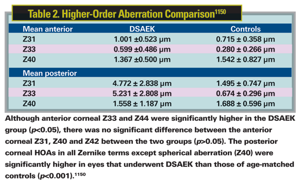
A group there performed DSAEK on 356 eyes, with 278 receiving donor grafts smaller than 8.5 mm and 78 receiving grafts of 8.5 mm or larger. Of those with a graft smaller than 8.5 mm, nine (3 percent) experienced graft dislocations; none developed primary graft failure. This was comparable (p=0.107) to the group of eyes receiving grafts 8.5 mm or larger with only two dislocations (2.6 percent) and no PGF. At six months, the mean BSCVA in the <8.5-mm group was 20/57 and in those with >8.5-mm grafts, it was 20/49 (p=0.313). All eyes with preop ocular comorbidities were excluded in assessing the postop VA. There was no significant difference in the hyperopic shift in the two groups. The only significant difference (p= 0.035) seen in the two groups at six months was in percentage of endothelial cell loss with the <8.5-mm donor graft group experiencing 34-percent ECD loss versus 26-percent for >8.5-mm grafts.
Percentage loss of ECD at six months was lower in the eyes receiving the larger donor grafts; this may be indicative of the larger number of endothelial cells delivered and lessened peripheral migration of endothelial cells over time in these eyes receiving the larger donor grafts, the researchers speculate. 2330
At
Average postop residual astigmatism was lower in the DSAEK eye compared to the PK eye by 2 D on manifest refraction and 7 D on keratometry. Average postop VA was better in the DSAEK eye as compared to the PK eye (logMAR .31 vs .43). On a subjective questionnaire, three patients reported visual acuity at six months to be better in the DSAEK eye, and four patients reported similar vision; no patients reported better vision in the PK eye. Additionally five of the seven patients reported more rapid recovery in the DSAEK eye; the other two patients reported the same speed of recovery for both eyes. Overall six of seven patients preferred the vision in their DSAEK eye, and one preferred the vision in the PK eye. Five of seven patients preferred the postoperative course of the DSAEK eye, one preferred the PK eye, and one noted a similar course with both surgeries. 1941
Avastin/Lucentis
Topical bevacizumab (Avastin), a recombinant humanized monoclonal IgG1 antibody that inhibits human vascular endothelial growth factor, has been considered for the treatment of corneal neovascularization. An investigation at Schepens Eye Research Institute and the Massachusetts Eye & Ear Infirmary,
In the study, bevacizumab 1% (10 mg/ml) was topically applied three times a day to the corneas of mice (n=30) divided into two groups: the first group with intact corneas and no obvious pathology or vascularization; the second group with corneal neovascularization induced by micropellet containing basic fibroblast growth factor. To evaluate corneal penetration in both groups, animals were euthanized at 12 and 24 hours, and three, five and seven days after the initiation of topical treatment for immunohistochemical analyses. Donkey anti-human IgG labeled with Cy3 was used for bevacizumab immunoreactivity detection.
Bevacizumab immunoreactivity was barely found in the corneal stroma in mice with intact corneas even after seven days of topical administration. Application of bevacizumab in mice with corneal neovascularization, however, showed high immunoreactivity in all layers of the cornea. 1488
Another group, this one at University of
Still another group at University of Erlangen-Nurnberg, in
At each visit, a routine Snellen acuity assessment, ophthalmic examination and fluorescein staining were performed. Corneal NV changes were assessed photographically. Seven patients (seven eyes) showed corneal epithelial defects during treatment, but six of these had preexisting epithelial defects. The one remaining eye had a persistent epithelial defect after pannus removal and limbal stem cell transplantation. In all other patients, no new epithelial defects were observed. Corneal sensitivity was difficult to assess in most patients due to multiple previous corneal operations. None of the 26 patients complained about any drug-related ocular or systemic adverse events during follow-up. No allergic reactions were observed.1481
A separate study at the same institution reached a similar conclusion, this time in a mouse model too, but with the additional finding that short-term topical application of the same concentration of bevacizumab produced no significant side effects on corneal epithelial wound healing after corneal abrasion, nor any effect on normal corneal integrity. 1469
There is also an in vitro study suggesting that bevacizumab is not cytotoxic to human corneal fibroblast and epithelial cells nor to human umbilical vein endothelial cells, and that all three cell lines showed evidence of proliferation upon exposure to the drug. At the University of Florida, Jacksonville, human corneal epithelial (HCE), human corneal fibroblast (HCRF), and human umbilical vein endothelial (HUVEC) cell lines, representing cells likely to exposed in topical treatment were exposed to increasing doses (0.1, 1 and 2 mg/ml) of bevacizumab. Cytotoxic and proliferative effects of this treatment were assessed by quantifying the cell numbers by WST-1 and crystal violet staining. Cell death was also assessed by flow cytometry and fluorescent microscopy using propidium iodide staining.
There were no cytotoxic effects of bevacizumab seen in any of the three cell lines (p<0.05). Cytometry and microscopy supported these results. There was a dose-dependant effect on proliferation on all three cell lines. HCRF proliferation increased up to 26 percent (SE 3.7; p<0.001), HCE up to 24 percent (SE 4.5; p<0.005) and HUVEC up to 37 percent (SE 10.6; p<0.01) with 2 mg/ml bevacizumab treatment as assessed with WST-1. Crystal violet assessment showed HCRF proliferation increased up to 35 percent (SE 2.4; p<0.0001), HCE up to 45 percent (SE 5.4; p<0.001) and HUVEC up 8 percent (SE 2.3; p<0.01) with 2 mg/ml bevacizumab treatment. 1486
A combined Swedish/Argentine group that previously studied Suramin (20 mg/kg) and bevacizumab (5 mg/ kg) found that intravenous injected separately, the combination diminished corneal NV. They're now evaluating a possible synergistic effect of the two drugs.
They induced corneal NV in two groups of nine rabbits. Group one was treated after injury with intravenous Suramab at a dose equivalent to 3 mg/kg of bevacizumab and 10 mg/kg of Suramin. The control group received no treatment. Digital photographs were taken at days 9, 15, 21 and 35. NV index (NVI) was calculated using the jimage program. Neovessels formation was quantified and given a score from 0 to 4 to each quadrant according to the centripetal growth of the longest vessel. Nine days after injury, the NVI was much higher in the control group (mean 4.7) than in the treated group (mean 1.5). Suramab significantly reduced neovessel formation (mean 6.1) compared with control animals (mean 13) at 35 days (p<0.01).
The systemic application of Suramab reduced corneal neovascularization. Duration of the effect was longer than that previously obtained with higher monodoses of bevacizumab and Suramin. Results suggest the presence of pharmacological synergy, they conclude. 1491
Research at the
The device was used in combination with a suture pull-through technique without folding the graft or touching the corneal endothelium. It was tested using three human donor corneas and three whole cadaveric eyes that were not suitable for transplantation. Following this procedure, the graft tissue was stained with Trypan blue to indicate endothelial damage. All three grafts indicated minimal to no Trypan blue staining of the endothelium following this procedure. 2344
A second study at Devers concludes that patients with previous glaucoma filtering surgeries can safely undergo DSAEK. The retrospective case series included 22 eyes of 20 patients (16 trabeculectomies, six glaucoma drainage devices). Additional procedures performed included three anterior vitrectomies, two cataract extractions with IOL placement, one IOL exchange, two sutured IOLs, two iridoplasties and two tube revisions. Intraoperative complications included two compromised blebs and one loss of donor tissue due to traumatic manipulation. The average follow up was 260 days (r: 28 to 628). Postoperative complications included one dislocation, one decentered graft, one retinal detachment, two graft rejection episodes and one late endothelial failure. There were no primary graft failures. In four eyes, one or more new glaucoma drops were initiated postop. No additional glaucoma surgeries were performed postoperatively. The average VA at the last exam was 20/143, representing an improvement from preop (p<0.001); 21 of 22 grafts were clear at the last follow-up and no eye lost vision. The average ECD at six months was 1,820 (r: 1,126 to 2,500) and the average cell loss was 32 percent (r: 13 to 55 percent) (n=4).
Eyes with glaucoma filters showed a significant improvement in vision with no primary graft failures and reasonably low dislocation (4.5 percent) and graft decentration (4.5 percent) rates. The overall intraoperative complication rate of 14 percent may, in part, be explained by the complexity of surgical procedures performed with DSAEK. Function of glaucoma filters may be affected after DSAEK, with 18 percent of eyes requiring additional medical therapy postop, however no filters failed during the postop period. While these are difficult surgeries, posing multiple unique challenges, excellent outcomes comparable to those seen in less complex eyes can be achieved, the group says. 2333
A retrospective review at the
The overall incidence of new or worsened postop glaucoma was 17.2 percent, which is towards the low range reported for the incidence of glaucoma after PK (10 to 53 percent). Since all of the patients were placed on corticosteroids after DSAEK, a steroid-induced pressure elevation cannot be excluded. None of the cases in the series required surgical intervention. Postoperative glaucoma after DSAEK may be less severe than glaucoma after PK, for which surgical intervention is often required.2313
Long-term soft contact lens wear leads to a change of corneal elastic properties, according to a
Twenty-eight eyes of 14 normal subjects (no contact lens wear) and 36 eyes of 18 subjects wearing soft lenses for more than five years were evaluated. There were three groups: normal (controls); 26 eyes of 13 subjects one day after discontinuation of lens wear; and 10 eyes of five subjects more than two weeks after discontinuation of lens wear. The corneal biomechanical properties were measured with the Ocular Response Analyzer; the main outcome measures were corneal hysteresis (CH) and the corneal resistance factor (CRF). Central corneal thickness was obtained using a custom-built real time anterior segment OCT.
In the control group, the mean CH value was 11.17 ±1.14 mmHg and the mean CRF was 8.75 ±1.44 mmHg. One day after discontinuation of lens wear, CH (10.25 ±1.05 mmHg) and CRF (7.47 ±1.17 mmHg) decreased significantly. In the two weeks-plus discontinuation of lens wear group, CH was 10.01 ±0.83 mmHg and CRF was 7.66 ±1.54 mmHg. There were statistically significant differences in CH and CRF between the control group and the one-day group. There were statistically significant differences in CH (p=0.006) and CRF (p<0.05) between controls and the two-weeks-plus group, but no significant differences between the one-day group and two-weeks group, and there were no significant differences in CCT (p>0.05) in three groups. There were significant correlations between CCT and CH (p<0.05) in three groups.661
A group at King's College London and
Using a 60-kHz femtosecond laser, they created 90- and 160-µm lamellar flaps, circular trephines and tangential delamination incisions in organ-cultured human corneas. Strain was measured before, immediately following and one week after surgery by interferometer.
Flap creation resulted in an increase in corneal strain in both the central and peripheral cornea (p<0.05). A similar magnitude of strain increase was associated with side-cut creation, but subsurface delamination did not cause significant strain change. The magnitude of strain increase correlated positively with vertical incision depth and did not recover during wound healing. Compared to 90 µm depth flaps, 160 µm flaps resulted in significantly less strain increase (10 percent versus 33 percent, p<0.05).
This means that central corneal elasticity increases following lamellar flap creation, challenging the belief that the central corneal flattening associated with LASIK flap creation reflects the cornea's primary mechanical response. The outward force conjectured to cause this flattening is a mechanical impossibility, they say. Instead, they suggest that the mechanical response of all corneas to keratorefractive surgery is steepening but changes in stromal hydration result in the flattening and hyperopic shift seen clinically. Keratectasia develops when creep and fatigue damage caused by IOP and its pulse accumulate more rapidly than can be repaired. Individual susceptibility is determined by genetic, mechanical, structural and cellular factors. They propose that this model canresolve many of the enigmas and apparent paradoxes surrounding ectasia development, including the regression of ectasia manifest as keratometric flattening seen following corneal collagen cross linking treatment.662
Dry Eye/Tears
If Dry Eye Syndrome is not further classified into preferentially evaporative or aqueous-deficient dry eye, the assumed diagnosis is usually the latter. This may lead to a misdiagnosis resulting in the recommendation of artificial tears, which usually fails in the most frequent cause of evaporative dry eye, meibomian gland disfunction. The assumption that DE-related symptoms usually correspond to aqueous-deficient dry eye may not be correct and may explain the failure of artificial tears in some patients. In a retrospective analysis at the University of Valladolid in Spain, researchers retrieved data on 271 patients (60 males, 211 females) with a mean age of 52.1 years (SD 14.5; range, 16 to 86) patients referred with chronic DE-related symptoms. When DES was diagnosed, it was subsequently classified into preferentially evaporative (pEV-DES, either due to MGD or other causes) or preferentially aqueous-deficient dry eye (pAD-DES).
Most patients (84.1 percent) had no diagnosis at referral; the remaining had a previous diagnosis of possible DES, blepharitis, and/or allergy. The time from onset of symptoms until the researchers' final diagnosis was 56.9 (SD 86.6; range, six to 516) months, and over 12 months in 68.3 percent of patients. The three most frequently reported symptoms were burning, foreign body sensation, and redness; chief complaint was foreign-body sensation, regardless of the final diagnosis. Redness-dryness, and eyelid itching-pain were the second and third most frequent symptoms in pEV-DES and pAD-DES, respectively. All patients had a final diagnosis of DES: 82.7 percent pEV-DES (all MGD) and 17.3 percent pAD-DES (29.8 percent Sjögren and 70.2 percent non-Sjögren). Allergy, rosacea and rheumatoid arthritis were the most frequently associated diseases. Low tear lysozyme level was preferentially found in pAD-DES (61.7 vs. 15.4 percent) and was positively correlated with meniscus height, breakup time, Schirmer test and normal conjunctival impression cytology. Patients with undiagnosed chronic DE-symptoms were more likely to have a final diagnosis of blepharitis (MGD). 2371
A group in
They used a Tearscope lighting system to assess tear-film dynamics (non-invasive breakup time) and lipid characteristics (thickness and contamination) in 160 non-presbyopes (age 28.1 ±7.3 years, r: 18 to 44) and 58 presbyopes (54.1±7.7 years, r: 45 to 83). The tear-film stability was mostly influenced by age; the breakup time was statistically significantly shorter for presbyopes than non-presbyopes (20.0 ± 13.9 vs. 13.2± 11.4 sec p<0.001) but no difference was found between gender (p=0.115). The lipid layer was significantly thinner (p=0.013) in the presbyopic population with a very marked synergic effect of age and gender. The lipid layer of presbyopic females was significantly thinner than that of presbyopic males (p=0.034) and than that of non-presbyopic females (p=0.002).
The aging tear film is characterized by significant changes in the tear lipid layer producing less protection from evaporation in the presbyopic population; the changes are more marked in women than men, the group says. The findings have significant implications for the management of presbyopic dry-eye sufferers, for whom treatment of decreasing evaporative problems is essential. 1541
Researchers at
Dr. Afshari is an associate professor of ophthalmology at the