Presentation
A 10-year-old Caucasian male presented to the Wills emergency room with a chief complaint of double vision. Five days prior to the onset of visual symptoms, his mother noticed nasal redness in both of his eyes. Three days prior to presentation, the patient developed constant binocular horizontal diplopia that was worst in left gaze. He also reported pain with extraocular movements and a headache.
Medical History
The patient had no significant past medical or surgical history, and he wasn’t taking any medications. Family history was notable for multiple family members with hyperthyroidism and hypothyroidism, including a grandfather with Graves’ disease. Social history was non-contributory. The review of systems was significant for weight gain over the past year.
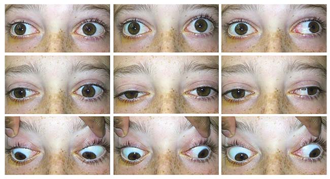 |
| Figure 1. Extraocular motility demonstrating limitations in adduction, abduction and elevation in both eyes. |
Examination
Ocular examination revealed a visual acuity of 20/20 OU. Pupils and confrontation visual fields were normal OU. Intraocular pressures were 17 mmHg OD and 18 mmHg OS. Extraocular motility of the right eye was notable for 20-percent adduction, 40-percent abduction, 30-percent elevation, and 100-percent depression. The left eye had 40-percent adduction, 90-percent abduction, 30-percent elevation and 100-percent depression (Figure 1). Ishihara color plates were 11/11 OU. External examination demonstrated 3 mm of proptosis of the right globe measured by Hertel exophthalmometry, with both globes exhibiting resistance to retropulsion. Right upper eyelid ptosis was present, and hypesthesia in the V1 and V2 dermatomes was absent. Anterior segment examination revealed nasal chemosis and injection OU but was otherwise unremarkable. Dilated fundus examination was normal OU.
What is your diagnosis? What further workup would you pursue?
Please click this link for diagnosis, workup, treatment and discussion.


