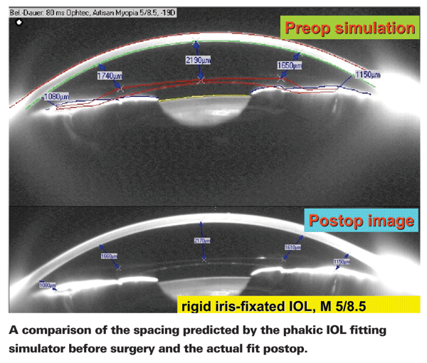
Although iris-fixated intraocular lenses have been in use for many years, they have always been associated with an element of uncertainty. A safe fit inside the eye depends on having sufficient space between the lens components and the surrounding ocular tissues, and those clearances are difficult to determine ahead of time.
Today, however, an optional software upgrade to the Pentacam HR (Oculus) is removing much of that uncertainty. As you know, the Pentacam can create a virtual 3-D model of the anterior chamber. The new phakic IOL fitting software preoperatively places an accurate 3-D model of the pIOL into the anterior chamber model and shows what the clearances will be between all parts of the pIOL and the surrounding tissues.
Avoiding Potential Problems
The idea of creating the phakic IOL fitting simulator came from Mana Tehrani, MD, who teaches ophthalmology at Johannes Gutenberg-University, in 
Dr. Tehrani explains that the need for the software arises from the limitations of a slit-lamp examination. "Through the slit lamp the anterior chamber depth may look OK, and you can measure the central chamber depth using ultrasound," she says. "If it meets the inclusion criteria, which is generally a depth greater than 3 mm, you may believe that the patient is a good candidate and proceed with surgery. But afterwards you may be surprised to find that the patient has pigment dispersion, or that there's contact between the haptics of the IOL and the endothelium. You may end up having to tell the patient that the lens must be explanted.
"The big advantage of this software is that it allows the surgeon to find candidates who are suitable for this surgery and eliminate poor candidates. This is mainly elective surgery, so it's even more important to exclude a patient who is likely to face complications or an explant."
The Simulator in Practice
"The Pentacam uses a rotating Scheimpflug camera to create a 3-D model of the anterior chamber," Dr. Tehrani explains. "The simulator software database contains all the biometric data you need to simulate any iris-fixated Artisan (Ophtec, Netherlands) or Verisyse (AMO) phakic IOL, including toric models. Once you enter the patient's refraction, the database gives you the refractive power of the pIOL that would be best to implant. The surgeon then selects the specific pIOL model and the software virtually fits the pIOL into the anterior chamber of that patient. It gives you the minimum clearances between all parts of the pIOL and the tissues surrounding them—the iris, the endothelium and the crystalline lens.
Until now, there was no way to determine precise clearances like this ahead of time. Dr. Tehrani says this gives the surgeon much greater security about immediate and long-term safety of the IOL being implanted.
"You can even rotate and shift the simulated pIOL manually to any position into which you are intending to later implant the pIOL," she continues. "The simulator software will let you see if the new position will still be safe. You can place the cursor on the simulated pIOL and manually shift it into different positions to see what the new clearances will be. The system is very flexible."
Dr. Tehrani notes that another advantage of the new option is that the surgeon can show the patient how the pIOL will lie in the eye after surgery. "Our patients are getting a much better understanding about the surgery we are planning to do. It makes all of this visual for them."
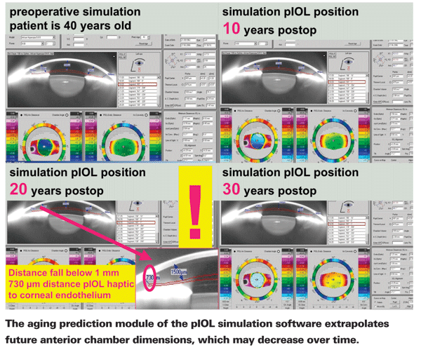
Another feature of note is the aging prediction module of the pIOL simulation software. This feature extrapolates the dimensions of the anterior chamber in the future, providing information about whether the pIOL is likely to become problematic over time. [See example, below.] "The anterior chamber decreases in size over time as part of the aging process," explains Dr. Tehrani. "This leads to a drop in the minimal distance between the implant and the lens and endothelium. The software uses an estimated amount of yearly anterior chamber size reduction to project what the clearances will be any number of years in the future. This is especially valuable in hyperopic patients who have very shallow anterior chambers."
Getting Even Closer
Dr. Tehrani admits that the software does have one or two limitations. "We did a study comparing the preop simulation with the actual postop position of the pIOL," she says. "We found a consistent deviation: The simulated pIOL was positioned up to 50 µm higher than the real one was after surgery. We realized this is because the virtual pIOL is not enclavated in the iris tissue. Unfortunately, there's no easy way to compensate for this, because every surgeon uses a different amount of tissue to enclavate the pIOL. So the actual lens will lie a little deeper than the simulation shows, and the surgeon should take that into account.
"The other limitation has to do with the aging feature," she continues. "We've discovered that the decrease in anterior chamber size over time may be different in different patients. The simulation uses 18 µm as the annual decrease, as suggested by Georges Baikoff.1 This number is pretty representative in Europe, but it's an average. We plan to conduct a study involving a large number of patients to refine this number so we can make a more precise prediction."
Dr. Tehrani says the Phakic IOL Fitting Simulation software is not included with most models of the Pentacam and must be purchased separately. However, it works with every available model, including the newest high-resolution device.
1. Baikoff G, Lutun E, Ferraz C, Wei J. Static and dynamic analysis of the anterior segment with optical coherence tomography. J Cataract Refract Surg 2004;30:9:1843-50.


