Any new device or technique that makes intravitreal injections simpler or safer is usually welcome. Two novel devices may help accomplish that goal—one by preventing contamination of the needle before the injection, the other by reducing the amount of equipment required during the procedure and standardizing the injection process.
Protecting the Needle
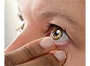
Alexander M. Eaton, MD, director of the Retina Health Center in Fort Myers and Naples, Fla., has helped develop a single-use “guarded injection device”—a tiny, collapsible sleeve that protects the needle from exposure to contaminants such as aerosolized saliva prior to and during the injection. “The needle comes with the sleeve already on it,” Dr. Eaton explains. “The surgeon draws the drug up with whatever needle he prefers; he then replaces it with the guarded needle for the injection.
|
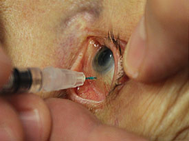
Dr. Eaton notes that the sleeve should eliminate the need for a lid speculum, particularly in patients who receive topical anesthetics. “When topicals are used you can never be completely certain that patients won’t squeeze or move when you give the injection. This device will prevent any contamination of the needle should that happen. This also has other benefits: Not needing a speculum reduces the amount of time required for the injection, and should also help reduce the risk of corneal abrasions and drying. Furthermore, it should save staff time because it eliminates the need to process and prepare the speculum.”
Dr. Eaton says he conducted a limited pilot study with 70 patients to compare the time required to do an injection in the traditional way (with a lid speculum) to the time required when the needle sleeve was used. “We measured the time from the instant the speculum or guarded needle was picked up to the moment the injection was completed and the instruments disposed of. Using the device, the injections took 23 ±8.2 seconds; done the traditional way, the injections took 34.4 ±5.7 seconds. The difference was highly statistically significant. We also measured the processing time for the speculum when it was used; that averaged 5 minutes and 28 seconds, which did not include autoclaving time.
“It’s worth noting that this timing was measured in a research setting with a trained assistant and all the equipment prepared,” he continues. “In real clinical use, the difference in time when using the sleeve may be even greater. Things are not always perfectly arranged when the surgeon enters the room.” Dr. Eaton notes that the data also showed a trend toward patients finding the guarded needle injections to be more comfortable than the traditional injections—due, at least in part, to not using a speculum.
Dr. Eaton acknowledges that it will be almost impossible to prove that the device reduces the rate of endophthalmitis, but he has done a study that suggests it might. “To test the effectiveness of the sleeve, we created ‘model eyes’ by filling sterile, empty Lucentis and Eyelea bottles with broth,” he explains. “Then we contaminated the outside of protected and unprotected needles with saliva and inserted them into the bottles.
“The bottles that were injected with unprotected needles all developed bacterial growth within 72 hours, while none of the others did,” he notes. “All the bacteria were in the Streptococcus viridans group—the most common cause of strep infection following intravitreal injections. Streptococcus is very virulent, which is why the visual outcomes with post-intravitreal-injection endophthalmitis have generally been poor.”
In terms of downsides, Dr. Eaton says adding the sleeve to the needles will undoubtedly increase the cost. “Our goal is to keep the cost difference very modest,” he says. “We hope to have the guarded needles available in the marketplace within six months to a year.”
Standardizing the Injection
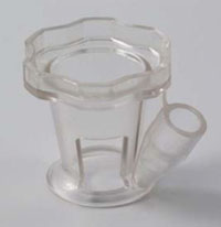
The disposable InVitria injection assistant (FCI Ophthalmics) is designed to help make intravitreal injections simpler, faster and more predictable. It consists of a polycarbonate shell that’s placed over the eye, with a guide tube into which the syringe is placed. A flange on the side of the device prevents any lashes from getting into the field, eliminating the need for a speculum.
|
Gokulan Ratnarajan, MBBS, who practices at The Royal Berkshire Hospital, Reading, U.K., has used the device for several months. (Neither he nor the institution has any financial interest in the device.) “When we were approached by the company to try the instrument it looked like a good idea, but we wanted to do a small pilot study,” he says. “So we compared 100 patients who received the injections by a conventional technique to 100 patients whose injections were performed with the aid of the device.”
In the study, researchers monitored patient pain scores, patient preference, surgeon perception regarding ease of insertion and the relative costs of the two approaches. The visual analogue pain scale score (from 0=no pain to 10=unbearable pain) in the conventional group was 2.58, compared to 1.38 in the InVitria group (p<0.01); the surgeon ranked the insertion of the device as easy in 89 cases, moderately difficult in 10 cases, and difficult in one case; and using the device cost about $12.35 less than using a conventional pack, which would amount to an annual savings of nearly $40,000 at the hospital.
“Patients liked the fact that they didn’t have a drape over their faces,” he notes. “And the hospital liked the fact that it was cheaper than the pre-made injection packs because less equipment, such as the surgical drape, calipers and disposable forceps, was required. Furthermore, in my experience, the learning curve for this device was simple and quick. I felt comfortable using it straightaway.”
Dr. Ratnarajan believes the device could be particularly useful if intravitreal injections are routinely given by health professionals other than the surgeon. “A device such as this might be less beneficial for an experienced surgeon, but for other health professionals or surgeons who are learning it could make injections safer and more consistent,” he says. “Either way, you still have the improved patient experience and cost savings.”
Regarding safety, Dr. Ratnarajan notes that their study is too small to draw any firm conclusions. “We didn’t have any major complications in either group, but you’d need a much bigger study to comment on that with any authority,” he says.
“Ultimately, we published our study in the British Journal of Ophthalmology,” he adds. “Our study does suggest that this instrument has value. We have now adopted it throughout the Institute and we’re very happy with it.” REVIEW