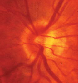
ASSESSING THE CONDITION OF THE OPTIC nerve and testing for functional deficits with a visual field are both essential when diagnosing and monitoring glaucoma. They may, however, present conflicting evidence; the optic disc may appear damaged while the visual field shows no functional loss, or the visual field may indicate a problem although no structural injury is apparent.
When that happens, it"s important to be aware that many different scenarios could explain the disagreement. Here, I"d like to review some of the possibilities we should take into account when we encounter such a disagreement during a patient"s first examination.
Disc Damage, Full Function
If the optic disc appears to be damaged but the visual field results are normal, there are three possible explanations:
- Prefunctional disease. Fields often fail to reveal early glaucoma because the visual system is highly redundant. Our retinas contain a variety of types of ganglion cells, any of which can pick up a white on white stimulus. Consequently, conventional visual field testing techniques only reveal ganglion damage when a substantial majority of the ganglion cells has been lost. (Specialized perimetry may have the most clinical value in this situation.)
- Normal anatomic variability. Another possibility is that the appearance of the optic nerve is misleading because of the relationship between the area of the optic disc and the volume of the neuroretinal rim. A large optic disc may have the same rim area as a very small optic disc, but the optic nerve can appear much more glaucomatous because the cup looks bigger.
Function Loss, Little Visible Damage
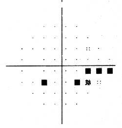
When the visual field shows loss of function but the structural damage appears minimal or nonexistent, at least three explanations are possible: The visual field results could be unreliable; disc damage could be more serious than it appears; or a nonglaucomatous process could be causing the functional loss.
When the visual field shows loss of function but the structural damage appears minimal or nonexistent, at least three explanations are possible: The visual field results could be unreliable; disc damage could be more serious than it appears; or a nonglaucomatous process could be causing the functional loss.
- Visual field testing error. Visual field test results can be inaccurate as a result of using the wrong refraction, patient fatigue, patient experience with the test, and a variety of well-described artifacts such as lens rim effects and field constriction because of small pupils (as a result of pilocarpine use, for example).
500 µm in diameter.
|
|
|
|