Carol L. Shields, MD, Miguel Materin, MD, John Epstein, MD, Jerry A. Shields, MD, Philadelphia
As a first year ophthalmology resident, each of us was trained to view the ocular fundus with the indirect ophthalmoscope and diagram the findings with colored pencils on a large retinal drawing sheet. This technique stimulated us to be comprehensive in our evaluation of the patient but also trained us to be exact. With time, our skills in fundus drawing improved to the level that we could create a fairly accurate depiction of the ocular fundus with simple pencils and paper.
This skill, however, is currently threatened by the development of high-resolution, wide-angle fundus photography using systems such as Retcam 120, Panoret-1000 and Optomap. These systems provide wide-angle images of the fundus without the need to superimpose photographs or create a montage of photographs. These wide-angle images achieve an accurate representation of the ocular fundus, perhaps replacing our fundus-drawing skills.
 |
 |
| Figure 1. A "homemade" camera fashioned from a Canon PowerShot G1 with light source bolted near the central axis of the lens. Above, the camera setup and at right, the fundus image. |
When assessing commercially available fundus camera systems, realize that there are several options. There are small-angle and wide-angle cameras (See Figure 2). There are cameras that utilize transpupillary illumination of the fundus and those that utilize transscleral illumination. There are those that are in contact with the cornea and others that are noncontact systems. And finally, most systems are stationary, but some of the newer systems are portable. These variables allow for the diverse capabilities of the several available fundus camera systems.


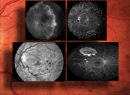
Figure 2. Available small angle and wide-angle cameras. A, The small-angle camera from Topcon. B, The wide-angle Retcam 120 camera. C, the wide-angle Panoret-1000 camera. (Not shown but available is a wide-angle camera from Optomap.)
Small-angle
The most popular fundus camera systems are the nonportable small-angle cameras. They provide high-resolution photography of the posterior pole of the eye and they image a small angle of the fundus, approximately 26, 45, 65 and 80 degrees of the posterior segment as measured with the vertex of the angle from the center of the vitreous cavity (See Figure 3).
 |
 |
| Figure 3. Fundus images with the various camera systems. A. Small angle camera image of small choroidal melanoma with serous retinal detachment showing 50 degrees of the fundus. B. Retcam 120 image of retinoblastoma showing 12 degrees of the fundus. | |
The small-angle systems typically have a light source mounted near the camera and employ transpupillary illumination of the fundus, in which the light is directed through the pupil into the posterior segment of the eye. Macular and paramacular images are excellent with such systems, providing high-resolution images of fine detail. These systems are ideal for the general ophthalmologist or specialist who wishes to document optic disc or macular detail.
The drawbacks of these small-angle cameras, however, are their inability to adequately image anterior to the equator of the eye and their inherent difficulty with imaging large fundus abnormalities (See Figure 4). In such cases, superimposing images can provide a general, but often piecemeal and inadequate picture of the large fundus abnormality.


Figure 4. Comparison of small-angle camera to wide-angle camera in a patient with choroidal melanoma. A. Small-angle camera image of choroidal melanoma. B. Wide-angle Panoret-1000 image of same patient as in 4A with choroidal melanoma.
Wide-angle
In order to document larger portions of the fundus in one image, wide-angle systems have been devised. The benefits of the wide-angle imaging systems include a panoramic view of the fundus and concomitant full perspective of the ocular condition, including associated findings elsewhere, remote from the area of concern. Additionally, wide-angle systems allow for photographic capture of the peripheral fundus, even into the pars plana in some instances. The potential disadvantages of the wide-angle systems include loss of fine detail and aberration from placing a concave view on a flat sheet of photographic paper.
The available systems are Retcam 120, Panoret-1000 and Optomap (See Tables 1,2). Retcam 120 is a contact system using transpupillary illumination to capture digital images of the fundus. Retcam 120 can provide high-resolution images for up to 120 degrees of the ocular fundus. This system is most commonly employed for young patients under anesthesia, such as those with retinoblastoma, retinopathy of prematurity and congenital ocular problems. We have employed Retcam 120 for more than 5,000 pediatric fundus evaluations and have been quite pleased with image quality.
| Table 1. Wide-angle Fundus Imaging Systems | |||
| Parameters | Camera systems | ||
| Retcam 120 | Optomap | Panoret-1000 | |
| Fundus illumination | transpupillary | transpupillary | transscleral |
| Camera type | contact | noncontact | contact* |
| Degrees of fundus imaged | 120 | 200 | 130 |
| Image resolution | good | skewed | good |
| Camera portability | yes | no | yes |
| Use for children | yes | sometimes | yes |
| Use for adults | no | yes | yes |
| Fluorescein angiography | yes | no | no** |
| Indocyanine green angiography | no | no | no** |
| *Good quality photos can be achieved with minimal contact to the coupling gel | |||
| Plans for angiography in the near future | |||
Retcam 120 requires wide dilation of the pupil, a clear crystalline lens and minimal media opacity. Imaging through intermediate or small pupil size, corneal opacity, lens opacity, pseudophakic lens or other media opacity yields suboptimal images due to the inability to illuminate the interior of the globe or difficulties with light reflection off irregular or artificial surfaces. Thus, Retcam 120 is generally limited to children without media opacity for the best resolution images and is not normally suitable for older adults. An outstanding feature of Retcam 120 is the ability to capture images of wide-angle fluorescein angiography, a feature not yet available on other wide-angle systems.
| Table 2. Wide-angle Imaging in Special Circumstances | ||
| Circumstances | Camera systems | |
| Retcam 120 | Panoret-1000 | |
| Small pupil | poor | fair to good |
| Cataract | poor | fair to good |
| Media opacity | poor | fair to good |
| Dark uvea | fair | fair |
Optomap is a noncontact scanning laser system using transpupillary illumination. This system is easy to administer as the patient places his or her chin on a chin rest, focuses on a spot directly ahead, and presses a button to take his/her own fundus image. Optomap claims to image up to 200 degrees of the fundus, but as anticipated, there is some inherent distortion in such an image.
Additionally, Optomap requires a cooperative, alert patient to take the photograph. This system might serve best as a screening tool as the image capture is broad, but the image quality suffers from aberration.
Panoret-1000 differs from all other wide-angle imaging systems in that it employs transscleral rather than transpupillary illumination. The idea for transscleral illumination was borrowed from the "Equator Plus" camera popularized by Oleg Pomerantzeff, MD, in the early 1970s.1-4 The Pomerantzeff film camera was the first to separate the illumination source from the camera and place it as a fiber optic source on the globe to illuminate the interior of the eye. Using a contact lens, the Equator Plus camera could photograph a field of approximately 148 degrees from equator to equator, and a little beyond. Satisfactory photographs with adequate resolution were obtained in most eyes imaged in this fashion and there were no local ocular complications.2 The Pomerantzeff camera was a breakthrough in fundus wide-angle photography, but fine details of the fundus in some cases were limited, mostly due to the brilliance of light at the site of scleral illumination.
Panoret-1000 differs from the Equator Plus camera in that it is a digital rather than film camera. Additionally, Panoret-1000 provides excellent image resolution to 20 µm of the retina with little scleral brightness at the illumination region.
Panoret-1000 can image approximately 130 degrees of the fundus using a 1024 x 1024 pixels CCD (charged coupled device) camera, acquiring images at a video rate for focus and alignment and saves high quality images at a rate of three frames per second, with a potential capability in the near future for wide-angle fluorescein angiography and indocyanine green angiography.
Like Retcam 120, Panoret-1000 is portable, but Panoret-1000 has the additional capability of imaging both children and adults as the transcleral light provides adequate illumination in patients with small pupils, cataract, pseudophakia and some corneal and media opacities. This system is ideal for imaging broad areas of the fundus for conditions such as diabetic retinopathy, retinal detachment, retinitis pigmentosa, peripheral retinal abnormalities and intraocular tumors.
Panoret-1000 can capture nearly the entire postequatorial fundus in one image. By manipulating the angle slightly, a clear image from the optic disc to the ora serrata can be achieved in any quadrant. Additionally, plans for adapting the camera with wide-angle fluorescein angiography and indocyanine green angiography are under way. We have used Panoret-1000 for more than 1,200 patients and have been pleased with its excellent image quality and ease of use.
We have observed no corneal or other ocular complications. A qualified examiner is necessary, however, to balance the light source, camera and foot pedal to properly capture an image. The main disadvantage of this system is that light transmission is less through more darkly pigmented uvea, a feature that is currently being improved.
Imaging of the ocular fundus has reached a level of excellence for both small-angle and wide-angle camera systems. There are advantages and disadvantages of both. Don't throw away your fundus drawings and colored pencils yet, as they will certainly complement the findings captured by the wide-angle camera systems.
The authors are from the Ocular Oncology Service at Wills Eye Hospital, 840 Walnut St., Philadelphia, PA 19107. They have no proprietary or financial interest in these devices.
Support was provided by the Eye Tumor Research Foundation, Philadelphia, PA (CLS), the Macula Foundation, New York, NY (CLS), the Rosenthal Award of the Macula Society (CLS), and the Paul Kayser International Award of Merit in Retina Research, Houston, Texas (JAS).
1. Pomerantzeff O. Equator-plus camera. Invest Ophthalmol 1975;4:401-6.
2. Ducrey N, Pomerantzeff O, Schepens Cl, Delori FC, Schneider J. Clinical trials with the equator-plus camera. Am J Ophthalmol 1977; 84:840-6.
3. Pomerantzeff O, Webb RH, Delori FC. Image formation in fundus cameras. Invest Ophthalmol Vis Sci 1979;18:630-7.
4. Pomerantzeff O. Wide-angle noncontact and small-angle contact cameras. Invest Ophthalmol Vis Sci 1980;19:973-9.