and Chirag P. Shah, MD, MPH
Presentation
An asymptomatic 49-year-old Bangladeshi female presented to the Wills Eye Institute to establish primary eye care. She had a history of "choroiditis" diagnosed by an outside ophthalmologist six months before presentation and was treated with laser photocoagulation.
Medical History
The patient's past medical history was significant for hypercholesterolemia and a seizure disorder. She had no history of diabetes or hypertension. Her medications included rosuvastatin and primidone. She had no past surgical history. The patient denied tobacco, alcohol or illicit drug use. Her family history was noncontributory. Her review of systems was negative.
Examination
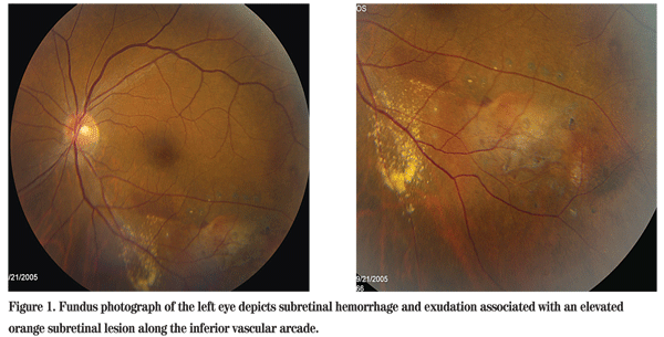
Best-corrected visual acuity was 20/30 in the right eye and 20/25 in the left eye. Intraocular pressures and anterior segment examinations were normal. Dilated fundus examination of the left eye was significant for subretinal heme, exudates and an elevated orange subretinal lesion along the inferotemporal vascular arcade (See Figure 1).

Diagnosis, Workup and Treatment
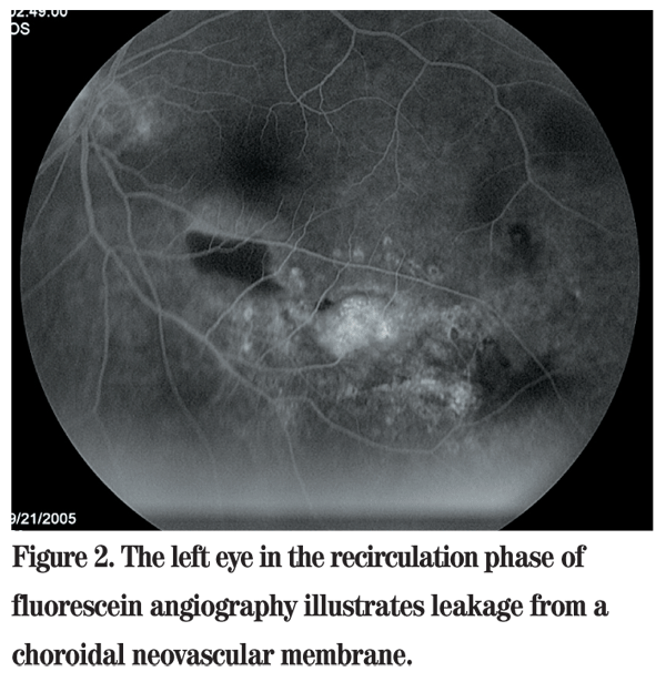
An intravenous fluorescein angiography and was first performed, which demonstrated leakage from a choroidal neovascular membrane (See Figure 2). The differential diagnosis of choroidal neovascularization in a 49-year-old Bangladeshi female includes polypoidal choroidal vasculopathy, age-related macular degeneration, pathologic myopia, angioid streaks, presumed ocular histoplasmosis syndrome and idiopathic CNV.
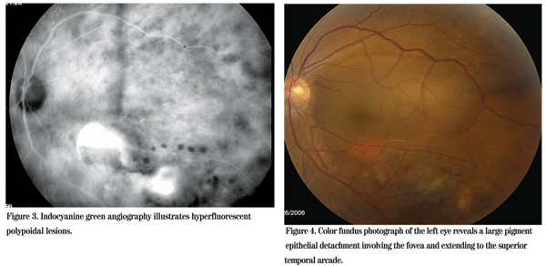
The orange choroidal lesion noted on clinical examination raised suspicion for PCV, particularly in a young Bangladeshi female with a normal fellow eye. Indocyanine green angiography was performed, which illustrated polypoidal choroidal lesions and confirmed the diagnosis (See Figure 3).

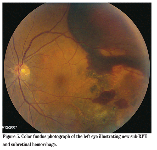
We discussed treatment options with the patient. She was asymptomatic and elected to monitor her vision with an Amsler grid and return regularly for retinal examination. She remained stable for 10 months, at which time she noticed distortion in her left eye. Visual acuity dropped to 20/60, with examination revealing a new pigment epithelial detachmentin the left eye (See Figure 4).

After a discussion with the patient regarding her treatment options and the known risks, she elected to receive intravitreal bevacizumab treatment. After two intravitreal bevacizumab (Avastin) injections, she had no improvement and her vision was 20/70 in the left eye. At that point, she underwent photodynamic therapy with marked improvement in the pigment epithelial detachment and her visual symptoms. Her visual acuity improved to 20/30.
Seven months later, she returned with new floaters and a cloud over her vision in the left eye. Her VA was 20/40. Dilated fundus exam and IVFA revealed new sub-RPE and subretinal hemorrhage along the superotemporal arcade, sparing the fovea (See Figure 5). She has since received multiple bevacizumab injections and PDT treatments.

Discussion
Polypoidal choroidal vasculopathy is an idiopathic condition characterized by orange lesions composed of dilated polyp-like clusters of abnormal vessels. The abnormalities typically lie in the inner choroid but external to the choriocapillaris. Afflicted patients develop a unique variant of CNV with associated complications, including hemorrhage, and serous retinal and pigment epithelial detachments.
Clinically, PCV resembles AMD; however, several characteristics can help distinguish between the two distinct clinical entities. PCV lesions have a higher likelihood of peripapillary involvement than AMD lesions and are less likely to involve the macula.
Macular, extramacular and peripheral variants of PCV have, however, been described.
PCV patients classically are non- Caucasian and present at a younger age with unilateral disease. The vasculopathy was originally described in African-American women, but more recent studies have confirmed a high incidence in both Chinese and Japanese patients as well. In contrast, AMD tends to affect older patients, most commonly Caucasians. ARMD is marked by drusen, retinal pigment epithelium alterations and exudative choroidal neovascular membranes. PCV patients often have a notable lack of associated drusen in either eye. PCV is, however, a protean disease and presents in all races, in both sexes, in all age groups and with marked variance in severity.
IVFA can often illustrate polypoidal lesions; however, ICG highlights the choroidal circulation and illustrates polypoidal lesions more effectively. The concurrent use of both studies along with the clinical picture distinguishes PCV from other disease entities.
Treatment for PCV is evolving. Photodynamic therapy effectively treats polypoidal lesions; but, as in our patient, recurrences are common. Anti-VEGF agents stabilize vision and reduce risk of exudative retinal detachment, but appear to have limited effectiveness in eliminating polypoidal lesions. Combination treatment for PCV with PDT and anti-VEGF agents may prove more effective than either treatment alone.
1. Yannuzzi LA, Dorik WK, Sforzolini BS et al. Polypoidal choroidal vasculopathy and neovascularized age-related macular degeneration. Arch Ophthalmol 1999;117:1503-1510.
2. Moorthy RS, Lyon AT, Rabb MF et al. Idiopathic polypoidal choroidal vasculopathy of the macula. Ophthalmology 1998;105:1380-1385.
3. Yuzawa M, Mori R, Kawamura A. The origins of polypoidal choroidal vasculopathy. Br J Ophthalmol 2005;89:602-607.
4. Ahuja RM, Stanga PE, Vingerling JR, et al. Polypoidal choroidal vasculopathy in exudative and haemorrhagic pigment epithelial detachments. Br J Ophthalmol 2000;84:479-484.
5. Sho K, Takahashi K, Yamada H, et al. Polypoidal choroidal vasculopathy: Incidence, demographic features, and clinical characteristics. Arch Opthalmol 2003;121:1392-1396.
6. Rosa RH, Davis JL, Eifrig CW. Clinicopathologic correlation of idiopathic polypoidal choroidal vasculopathy. Arch Opthalmol 2002;120: 502-508.
7. Lai TY, Chan WM, Liu DT, Luk FO, Lam DS. Intravitreal bevacizumab (Avastin) with or without photodynamic therapy for the treatment of polypoidal choroidal vasculopathy. Br J Ophthalmol 2008;92:661-6. Epub 2008 Mar 20.s.


