Presentation
A 78-year-old male presented to the Wills Eye Ocular Oncology Service with a chief complaint of pain, decreased vision and fullness around his left eye. He had initially presented to his primary ophthalmologist with this complaint five months prior and was placed on prednisolone acetate 1% four times daily without relief. He was then referred to a retina specialist, who diagnosed him with uveitis and hemorrhagic choroidal detachments. The retina specialist escalated his therapy to atropine 1% twice daily and prednisolone acetate 1% every hour. The patient’s pain was slightly improved. An attempt to drain the hemorrhagic choroidal detachment at that time was unsuccessful.
|
A limited workup of a prostate-specific antigen level and chest X-ray were within normal limits. The patient could not undergo magnetic resonance imaging due to his pacemaker, so a computed tomography scan of the orbits was performed, showing only vitreal hemorrhage without observable tumor. A contrast study could not be performed due to the patient’s severe renal impairment. B-scan ultrasound was also negative for tumor.
Medical History
Past medical history was significant for diabetes mellitus, congestive heart failure status post-pacemaker placement and end-stage renal failure. He was on numerous chronic medications for management of diabetes, hypertension and renal failure. Family history was noncontributory.
Examination
Ocular examination revealed a visual acuity of 20/25 in the right eye and light perception in the left eye. The right eye was unremarkable except for mild non-proliferative diabetic retinopathy.
The abnormal features were limited to the left eye. The pupil on the left was irregular and nonreactive with a relative afferent pupillary defect. Visual fields testing was limited by the poor visual acuity. Extraocular motility was limited by the mass lesion. An accurate intraocular pressure could not be obtained by Goldmann applanation or by Tono-pen.
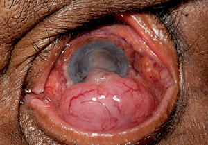
External exam revealed an epibulbar mass measuring 11 mm long, 14 mm wide, and 5 mm thick on the left eye. The mass appeared to be extending from the sclera (See Figure 1).
Slit lamp examination of the left eye was notable for a large epibulbar mass that appeared to have internal vessels, as well as adjacent conjunctival vessels. A sentinel vessel was also present. There was corneal edema and neovascularization of the iris. The anterior chamber appeared shallow, and there was a posterior chamber intraocular lens in place.
There was no view posteriorly on the left.
What is your differential diagnosis? What further workup would you pursue?


