SLIT-LAMP BIOMICROSCOPY IS THE STANDARD WAY TO EVALUATE donor corneas. However, some corneal scars that would make a cornea unsuitable for transplantation, such a LASIK flap, can be hard to detect with the slit beam. That's why Erkin Abdullayev, MD, G. Scott Bryan, and their co-workers at the Wichita Eye Foundation/Mid-continent Eye Bank are developing a system of backlighting that makes corneal features stand out more prominently. Here's how the system works.
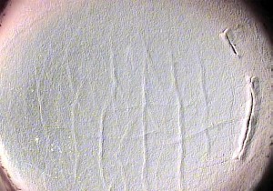 |
| Corneal Backlight Microscopy uses two lights to retroilluminate donor corneas to delineate such features as scars and folds. |
Dr. Abdullayev believes that it's easier to see corneal features during a slit-lamp evaluation of a live eye because the light from the slit beam is reflected back to the observer by the retina and iris. With a donor cornea, however, these structures are no longer there, so an evaluator, usually a technician, has to sometimes struggle to discern scars or other abnormalities.
To help offset this problem, Dr. Abdullayev places two lights behind the cornea, which is in a plastic viewing chamber in front of the slit-lamp microscope. He says the first light, a small 40-watt bulb, gives the observer the general image of the whole cornea, "like seeing the Earth from space," he says. "You see all the layers of the cornea, just like you'd see the upper and lower atmosphere of the Earth, with the 'clouds' in this case being signs of trauma or disease." This light is located 1 cm to 5.25 cm behind the cornea. The second light is located 7 or 8 inches behind the viewing chamber, and helps create an artificial red reflex that improves the imaging of stromal scars and previous surgical cuts. The evaluator still uses the slit lamp to confirm the findings, since it can give a 3-D view to show which feature lies above another.
The other important aspect of their system is a digital camera, mounted on the slit lamp and connected to a high-resolution 17-inch monitor. "As opposed to the slit lamp, where you use only the microscope and have to move the slit beam over the cornea, with the backlit image on the monitor, you can see the whole image," explains Dr. Abdullayev.
He says using a monitor allows the tech to digitally measure the image and place circular overlays on it that can tell him if any abnormalities he sees would be within the diameter of the donor corneal button. Without this, the technician would have to make cumbersome measurements using the slit-lamp beam and the slit lamp's adjustable ruler. Since the images are digital, they can also be sent to prospective surgeons, who can evaluate a cornea by looking at its actual image, rather than just relying on a written description of it.
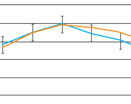
In a study presented at the 2004 American Academy of Ophthalmology meeting in New Orleans, Dr. Abdullayev and his colleagues used their backlight corneal microscopy method to evaluate 1,100 donor corneas, 17 of which had LASIK flaps. They used the slit lamp as a control. BCM detected an "obvious" flap edge in all the LASIK corneas, compared to only six (36 percent) caught by the slit lamp.
Though the BCM system is in the initial stages right now at the Mid-continent Eye Bank, Dr. Abdullayev says it's already increased his staff's abilities. "It's very easy, very fast and very informative," he says.
For more information on the BCM system, contact Dr. Abdullayev at eabdullayev@mid-continent.org.