A 9-day-old male infant was noted to have a “cloudy left eye” while in the postpartum nursery. The baby was otherwise healthy following delivery by repeat cesarean section at 36.5 weeks.
Medical History
Routine prenatal ultrasound suggested an absent corpus callosum which was confirmed with prenatal magnetic resonance imaging. Subsequent single nucleotide polymorphism chromosomal microarray was normal.
Examination
The patient was well-appearing. There was a large region of dermal melanocytosis over the scalp, but the head was normocephalic. His reflexes were intact and appropriate for his age. There were no focal neurologic signs.
On ocular examination, the child showed a preference for fixation with the right eye and a left exotropia but retained full extraocular movements without nystagmus. There was no afferent pupillary defect. The remainder of the external exam was unremarkable.
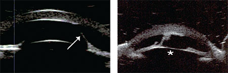 |
| Figure 1 (left). Ultrasound biomicroscopy of the right eye showing iridocorneal strands (arrow). Figure 2 (right). Ultrasound biomicroscopy of the left eye showing a slightly anteriorly displaced lens (asterisk) with a central scalloped defect of the posterior cornea. There are pronounced iridocorneal adhesions. |
Handheld slit lamp examination demonstrated a faint endothelial scar just above the visual axis of the right eye, without corneal edema. The left eye exhibited a full-thickness, white central circular scar. Both corneal diameters measured 9.25 mm. A single iridocorneal strand was noted inferiorly in the right eye, while the left eye had three strands to the scar. The anterior chamber was noted to be shallow in the left eye relative to the right. Dilated fundus exam of the right eye was unremarkable, and although the left provided a hazy and limited view, there were no obvious abnormalities.
Please click this link for diagnosis, workup, treatment and discussion.


