Determining when a patient has begun to develop glaucoma has always been problematic. Checking intraocular pressure is quick and easy, but not very accurate as a diagnostic tool, and doing a battery of more complex tests on every suspect is simply not practical.
Now, however, a new use of visual evoked potential technology called "isolated-check VEP," or icVEP—currently beginning Phase II clinical trials—is showing great promise as a potential diagnostic tool. And unlike multifocal VEP or standard visual field tests, this device has shown a remarkable ability to distinguish subjects with early glaucomatous damage from normals within seconds, because it only tests the most damage-sensitive neural pathway, and only tests it in the central retina. Its creators hope it will prove to be more objective than perimetry, and a useful tool for identifying and monitoring glaucoma patients.
Staying Centered
Vance Zemon, PhD, professor of psychology at the Ferkauf Graduate School, Albert Einstein College of Medicine of Yeshiva University in Bronx, N.Y., has worked with VEP technology for 30 years. He is largely responsible for developing the icVEP technology upon which the new device is based.
"The multifocal VEP technology that's currently available is appealing to scientists and clinicians because it gives you something akin to an electrophysiological visual field test," he explains. "The patient views patches of light and dark in a pseudo-random pattern while electrodes pick up the responses coming from the different parts of the retina. This allows you to evaluate the integrity of the retina much as a visual field test does, with the advantage that the subject doesn't have to make judgments.
"The problem," he continues, "is that multifocal VEP requires running very long sequences of stimuli, which can take 20 or 30 minutes for a complete test. It's not easy for patients, especially if they're elderly or haven't done it before. Also, the operator has to look at a printout of little waveforms and judge what's a true response and what's noise. The process is time-consuming and not 100-percent objective."
In contrast, icVEP tests only the central retina. "A colleague, Vivian C. Greenstein, PhD, tested patients with very specific stimuli such as pure color contrast," he says. "She found that—contrary to popular belief—central vision is affected in early glaucoma. The idea that this isn't the case probably came about because tools such as a standard visual field aren't particularly sensitive."
James C. Tsai, MD, professor and chair of the Department of Ophthalmology and Visual Science at Yale University School of Medicine in New Haven, Conn., has been involved with the icVEP instrument since the beginning of the Phase I studies. (He's the lead investigator for both clinical trials, but has no proprietary interest in the instrument.) "When we were approached about using this technology to help detect early glaucoma, we were really surprised that it was possible," he says. "The premise is that patients with glaucoma may have 20/20 vision according to Snellen visual acuity, but when you look at their contrast sensitivity curve they're abnormal compared to someone without glaucoma."
Dr. Tsai notes that this test was designed for patients with glaucoma who have relatively good central visual acuity—at least 20/30 or better—and who are able to focus on a central test object. "For that reason, unlike multifocal VEP technology, this won't be helpful for patients who have poor central vision," he says. "On the other hand, the beauty of the test is that each test recording takes only about two seconds. Once the electrodes are placed on the scalp, it's very quick data acquisition."
How icVEP Works
"The visual system is organized into parallel, independent streams of different kinds of information flowing from the retina," explains Dr. Zemon. "Different cell types are involved, and the different types don't talk to each other. Instead, they pass their signals on to other 'like' cells in the retina, and eventually to the primary visual cortex in the occipital lobe at the back of the brain.
"Researchers have discovered that there are two basic cell types originating in the retina at the very first synapse between photo-receptors and bipolar cells," he says. "One is a larger cell, which makes up what is called the magnocellular pathway because it runs to the magnocellular layers of the lateral geniculate nucleus in the middle of the brain—the major relay center. The retinal ganglion cells that feed this pathway are now called M cells. Similarly, the smaller retinal ganglion cells that feed the parvocellular pathway are called P cells. In addition, researchers have discovered a third pathway in the lateral geniculate nucleus called the koniocellular pathway. The cells making up this pathway seem to be critical for processing our blue/yellow color information, and that fact, along with their limited number, makes tests such as short wavelength automated perimetry (SWAP) possible.
"There are a number of important differences between the M cells and P cells," he continues. "The large M cells are very sensitive to low levels of luminance contrast; P cells are not. On the other hand, P cells are very responsive to color contrast, but M cells are not. Furthermore, M and P pathways are subdivided into 'on' and 'off' cells, which are devoted to relaying certain types of information.
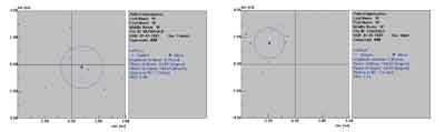
Representative icVEP test results, based on eight runs. If the circle includes the origin, the signal to noise ratio is less than 1 and the test is positive. If the circle does not include the origin, the signal to noise ratio is greater than 1 and the test is negative. Left: 15% bright-check condition for one glaucoma patient (SNR=0.32). Right: Same test condition for a representative control (SNR=1.85).
"With our VEP techniques, we developed stimuli that specifically draw a response from individual pathways," he says. "We discovered that a small sample of glaucoma patients showed significant disruption of the 'on' division of the magnocellular pathway that responds to bright, low-contrast information. Also, we realized that this damage was detectable in the foveal region, making it unnecessary to test a wider area of the retina. So when George Hu, a former student I'd worked with at Rockefeller University with a PhD in electrical and computer engineering, contacted me about developing an instrument that would detect early glaucoma, it seemed clear that this was the way to do it."
Taking the Test
After teaming up with Dr. Zemon, Dr. Hu developed the prototype of the icVEP device, with the intention of developing the technology into a marketable instrument for clinicians. The National Institutes of Health awarded them a Phase I Small Business Innovative Research grant, and they were able to test their prototype on a population of glaucoma patients, suspects and normals at Columbia University in New York.
Dr. Zemon describes how the version of the instrument used in the Phase I trials works (noting that the version being developed for use in Phase II will have several significant improvements). "Because the test requires the ability to focus on a target, it is designed for patients whose central vision is relatively intact, with 20/30 visual acuity or better," he says. "If you looked at their visual field tests, most glaucoma patients who fit this description would be classified as having mild glaucoma.
"After the patient is seated at the instrument, we put three electrodes on top of his head with a little water-soluble paste," he explains. "The patient is then instructed to look at a square field with a small fixation cross in it in the middle of the screen. The operator monitors the patient's gaze to ensure he's looking at the fixation object. When he is, the run is started with an auditory cue to alert the patient; then the cross gets smaller to hold his attention.
"The run itself last only two seconds," he continues. "The first second is an adaptation period, during which we don't collect data. Then the isolated-check pattern switches to double the contrast that was present during the adaptation period, for one second, and the data are collected. Because the test is done so quickly, we repeat this eight times for each eye."
Once the instrument has collected these eight EEG samples, according to Dr. Zemon, it first determines whether there's an outlier among them. If there is, it instructs the operator to do another run until it has eight consistent runs. Then the computer does an automatic multivariate statistical analysis of the data, producing a signal-to-noise ratio for that eye. If the ratio is greater than 1, it means there is a healthy response there, with a 95 percent confidence interval. If it finds a ratio equaling 1 or less, the eye has failed the test. "If the eye fails the test, we redo it to make sure the result is accurate," he notes. "If the eye fails twice, it indicates there's a problem that needs to be looked into."
Dr. Zemon adds that the instrument also has built-in artifact rejection features. "If too much electrical noise creeps into the recording, indicating that an electrode has come loose, for example, the instrument will automatically signal the operator that it is rejecting those data," he says.
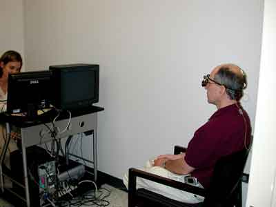
In the prototype model used in Phase I testing (above), the operator monitored the patient's gaze to ensure fixation. The next model will be largely automated and feature a chin rest and infrared fixation
Although the sample used in the Phase I trial was small, the results were impressive. "The accuracy we obtained in Phase I testing for that critical pathway, the magnocellular 'on' pathway, was just over 94 percent, much higher than any of the techniques that are currently out there," says Dr. Zemon. "The specificity of our test—one component of accuracy—turned out to be 100 percent; all normal subjects passed the test."
Taking the Next Step
Drs. Hu, Zemon and colleagues are now ready to begin a Phase II clinical trial which will run over a two-year period at multiple clinical sites, including the University of Alabama at Birmingham and the Hamilton Eye Institute at the University of Tennessee Health Science Center. "We want to measure data from a larger sample so we can get more accurate statistical data about sensitivity and specificity, and hopefully improve the quality of the instrument," says Dr. Hu. "Also, we'd like to test each patient several times, several weeks apart, to make sure the machine meets the repeatability requirement. We didn't do this in the Phase I trial, but we did do some repeatability testing with Prof. James Gordon at Hunter College of the City University of New York, and the results were very good. In Phase II, we'll do this kind of testing with every subject."
Dr. Hu has been upgrading the instrument's accuracy and ease of use in several ways for the next trial and for eventual marketing as a clinical tool. "Unlike the research version," notes Dr. Hu, "the current model is largely automated and more convenient for the user. It presents the stimulus to the patient and automatically collects and processes the data and generates results. Also, laboratory instruments using VEP traditionally have involved multiple measurements to allow for noise processing. Our device uses a shorter measurement period and processes the results using a statistical method that generates more stable and reliable data. As a result, we've reduced testing time and increased data reliability."
Dr. Zemon lists additional improvements. "In Phase I, the operator had to decide whether the patient was fixating carefully," he says. "Now we're building an infrared system that will facilitate monitoring fixation. We're enhancing the detection of alpha brain waves, which can increase the noise level, and we're adding a head and chin rest so the subject stays at a fixed distance. We're trying a higher resolution video board to see if it produces better responses. And, we're doing pilot testing to determine whether changing the stimulus slightly will enhance the accuracy of the results."
Reaching the Marketplace
"Ultimately, if testing proves it effective, we hope to manufacture an instrument of this kind," says Dr. Zemon. "Unlike the original research version, the newest version is very user-friendly. It's based on the Windows operating system, so it should be intuitive for anyone who uses a computer. Hopefully, once it's in the hands of clinicians and they're getting positive feedback, they'll want to continue to use it in their practice."
Dr. Hu says he hopes the instrument—tentatively named Neucodia—will be available for sale within six months. Asked about cost, Dr. Hu says their current idea is to simply lease it to clinicians at the outset, charging only a portion of the reimbursement provided by insurance for each use. (A CPT code for VEP testing already exists.) "This way the doctor doesn't need to worry about making an investment," he says. "We'd install the instrument in the office at no charge. If the doctor is satisfied, he can eventually purchase it; if not, we'll take it back. Also, we're trying to make the machine so easy to use that a technician or secretary can use it with minimal training, so the doctor can try it without investing money or time."
Regarding the future, Dr. Tsai says he's cautiously optimistic. "In our initial study, where we compared clear-cut examples of normal and glaucoma, it did very well," he says. "Time will tell where it turns out to be most useful. Today we rely on visual fields along with imaging technologies such OCT, HRT and GDx, for glaucoma diagnosis. But even with those, we often don't get enough information. This technology should add another piece to the puzzle."


