Presentation
 |
A 20-year-old Caucasian female was referred for evaluation of an enlarging left conjunctival lesion. One month prior to presentation the patient had noticed what she perceived to be a “popped blood vessel” in the conjunctiva of her left eye. She noted mild redness around the lesion, but denied pain or discharge. Vision was stable with glasses. There was no history of ocular trauma or contact lens wear. The review of systems was within normal limits. The lesion didn’t respond to topical tobramycin initiated by the referring ophthalmologist.
Medical History
The patient had no significant past medical or surgical history. Family history was positive for a thyroid disorder in her mother. The patient was a non-smoker, and denied illicit drug use. She reported having an allergy to doxycycline.
The patient’s medication list included pantoprazole 20 mg tablet two times per day and a combination oral contraceptive daily.
Examination
Best-corrected visual acuity was 20/25 in the right eye and 20/30+1 in the left. Pupils were equal, round and reactive bilaterally with no relative afferent pupillary defect. Intraocular pressure was 20 mmHg in both eyes. Visual fields were full to confrontation bilaterally. Extraocular motility was full bilaterally. There was no evidence of proptosis. Adnexa and eyelid examination was within normal limits bilaterally.
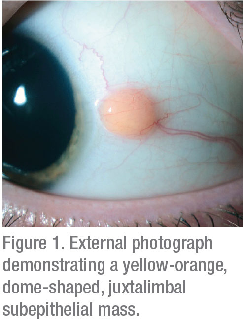
Slit lamp examination of the left eye demonstrated a circumscribed, soft, solid, yellow-orange, dome-shaped, temporal juxtalimbal subepithelial epibulbar nodule that measured 3 x 3 mm (Figure 1). A prominent vessel was noted temporal to the lesion. There were no associated cysts or pigmentation. The lesion was slightly mobile and did not appear to be completely adherent to the underlying sclera. The remainder of the anterior segment and funduscopic examination in both eyes was unremarkable.
What is your diagnosis? What further workup would you pursue? Please click this link for diagnosis, workup, treatment and discussion.


