LASIK is widely accepted as a safe and effective method for correcting refractive errors. An estimated 1.5 million annual LASIK procedures are performed worldwide.1 As a procedure is performed with increasing frequency, unforeseen events may occur when patients with unidentified contraindications undergo the surgery.
A thorough understanding of the potential complications of LASIK and various strategies to prevent and manage them is essential for the surgeon performing the procedure. Complications including corneal ectasia, flap problems and epithelial ingrowth may be avoided with careful preparation and attention to detail. This article will review the special challenges inherent in performing LASIK on corneas with unusual characteristics.
Steep Corneas
Presentation of abnormally steep corneas should raise a few important questions for the surgeon. The foremost significant risk factor to be ruled out in patients with steep keratometric readings is the presence of irregular astigmatism, which is a hallmark of keratoconus and form fruste keratoconus.2
A recent search of the peer-reviewed literature revealed preoperative topographic evidence of form fruste keratoconus in 13 of 27 cases of progressive and visually threatening ectasia following LASIK.3 Computer-assisted videokeratoscopes with color-coded maps and topographic indices are the most sensitive and sophisticated devices for confirming the diagnosis of keratoconus.
With such devices, keratoconus appears as an area of increased surface power surrounded by concentric zones of decreasing surface power. Three features are common to keratoconus videokeratographs that use sagittal topography: a localized area of increased surface power (46 D), asymmetry in the superior-inferior (S-I) power of greater than 1.50 D, and skewed steep radial axes above and below the horizontal meridians (>15 degrees).
Ultrasonic pachymetry may be useful to confirm corneal thinning in patients with suspected keratoconus on slit lamp examination or videokeratography. It cannot be solely relied on, however, to make the diagnosis because of the large range and variability of pachymetry readings both centrally and paracentrally in the normal population.3
Steep corneas have also been implicated in the etiology of thin, irregular or buttonhole flaps.4 The lamellar flap in LASIK should be deeper than Bowman's layer, sparing it from laser ablation. A flap is considered thin when the keratome cuts above or within the 12-µm thick Bowman's layer.1 Suspect a flap of less than 60 µm of being thin because the thickness of the corneal epithelium is approximately 50 µm.
 |
 |
 |
 |
| Topographic maps of a 26-year-old female suspicious for form fruste keratoconus in OS, who underwent bilateral LASIK and presented with inferior steepening in the eye one week following surgery. | |
A buttonhole in the flap occurs when the microkeratome blade travels too superficially and breaches the central epithelial/Bowman's complex. A mean keratometry reading of 46.70 D in patients who develop buttonholes has been reported,5 and another study found mean K's of 44.20 D to be significant.6 One possible hypothesis for buttonhole formation is that steep corneas buckle centrally upon applanating pressure, which results in a central dimple missed by the blade. Another hypothesis is that corneas with high keratometric values offer greater resistance to cutting when applanated, leading to anterior motion of the blade with respect to the cornea. Also, steeper corneas may also dry faster leading to corneal dessication, which plays a role in buttonhole formation.
The safest management of a thin, irregular or buttonholed flap is to reposition the flap and abort the procedure.1,4 Proceeding with laser ablation underneath the irregular flap may cause scarring, irregular astigmatism, epithelial ingrowth and loss of best corrected visual acuity. A thin flap is, however, difficult to reposition and more prone to striae.
A new flap may be recut 20 to 60 µm deeper than the initial cut. This can be attempted about 10 to 12 weeks later with a different microkeratome or a larger diameter flap size. However, prevention is the ideal solution.
In patients with steep corneas the microkeratome needs to be set to a deeper cutting depth, assuming that the corneal thickness is sufficient for the intended refractive correction. Meticulous maintenance of the microkeratomes and inspection for damage prior to each procedure is crucial. In addition, ensuring adequate suction with pressure above 80 mmHg is essential for safe flap creation. Pneumotonometer measurement is probably most reliable. Pseudosuction must be avoided. This is often caused by conjunctival clogging of the suction port which presents as a discrepancy between intraocular pressure and suction pressure.
Another factor to consider in the amount of treatment attempted in a steep cornea is the final postoperative keratometry. In our clinical experience, we feel that it is not safe to treat corneas that will have a postoperative keratometry > 50 D because this may lead to optical aberrations. For example, the maximum safe hyperopic treatment in a 46-D cornea would be +4 D.
Flat Corneas
Patients presenting for LASIK with excessively flat corneas with keratometric readings of < 42 D are at a higher risk for developing free caps, which are unintended complete dissections of the corneal flap.1 This occurs because less tissue may be brought forward into the zone to be cut by a microkeratome, increasing the possibility of a free cap.
A free cap does not prevent the surgeon from completing the case. If the diameter of the exposed stroma is equal to or larger than the intended optical zone, laser treatment can proceed while the cap is conserved in an anti-dessication chamber or on the conjunctiva with the epithelial side down during the photoablation.
After the laser ablation, the cap is repositioned in the same orientation as before surgery, and five to 10 minutes drying time allows the cap to adhere without sutures. The use of orientation marks is helpful in realigning the cap. A bandage contact lens helps to tamponade the cap and protect it from displacement by palpebral friction. If the cap can't be recovered, the epithelium is allowed to grow centrally in a manner similar to that after superficial keratectomy procedures. This may be associated with a significant hyperopic shift. We defer retreatment until refractive stability is achieved.
The postop keratometric outcome also guides the amount of treatment in a flat cornea. Most refractive surgeons feel that it is a good practice not to treat if the final result causes keratometry readings less than 35 D. Some feel comfortable in treating up to 30 D.
Irregular Corneas
Asymmetric bow-tie patterns or localized corneal steepness on topographies in patients not strictly meeting the criteria of keratoconus can also present in cases of contact lens warpage, especially in rigid contact lens wearers.
As a rule, patients should be out of soft contact lenses for at least three days and rigid gas permeable lenses for at least three weeks before preop assessment. Soft toric contact lenses seem to behave more like RGPs and their use should be discontinued accordingly. These guidelines represent a minimum.7
Another rule of thumb is to discontinue contact lenses for one month per decade of contact lens wear. Contact lenses can cause warpage, mask existing astigmatism or induce irregular corneal astigmatism. LASIK should not be planned until refraction and topography have stabilized and normalized completely. This may take months in long-term RGP wearers. In cases where topography does not normalize, consider custom cornea ablation.
In patients with astigmatism, the goal of treatment with LASIK is to bring the differing focal lines in the eye together on the retina. This can be done by flattening the steep meridian or steepening the flat meridian.8 When treating simple or compound myopic astigmatism, the excimer beam also flattens the steep axis by removing more tissue along a central band aligned along the prime axis, with progressively less ablation in the periphery. This doesn't affect the flat meridian.
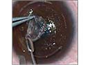
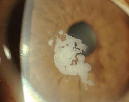
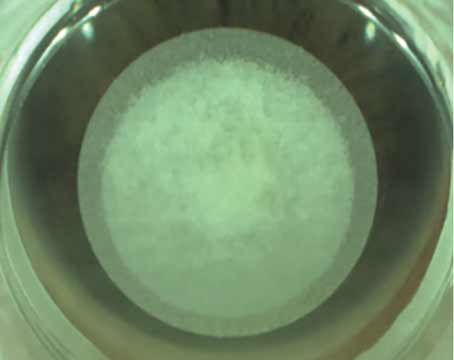
 |
| A LASIK flap with buttonhole |
An additional factor to be considered in the planning of an astigmatic procedure would be to vary the hinge placement based on the intended astigmatic correction. Because one meridian of the astigmatic laser treatment is longer than the other, varying the flap hinge placement either superiorly or nasally would allow more room for the long-axis ablation to take place. For example, in myopic with-the-rule astigmatism, the elliptical ablation would be longest in the horizontal direction, so a superior flap would best accommodate it.
Different algorithms are being developed for hyperopic cylinder to maximize refractive effect, minimize tissue ablation and mitigate regression.8 One common feature of toric treatments is elliptical ablation patterns. The smaller the optical zone, the higher the likelihood of postop haloes, glare and potential for decentration. In patients with large mesopic pupils and high astigmatism, the optical zone may be a limiting factor, especially if the patient has against-the-rule astigmatism, since most glare comes from a horizontally smaller zone.
Finally, in patients with irregular astigmatism, it is recommended to only treat with custom cornea ablation after keratoconus has been ruled out.
LASIK has a very high success rate. The demand for especially high standards of safety for this surgery is mandated because relatively healthy eyes are placed at risk every time the procedure is performed. These risks can be minimized by learning from experience, analyzing outcomes, and exploring new territory thoughtfully and ethically.
Dr. Krueger is medical director of refractive surgery and Dr. Waheed is a fellow in refractive surgery at the Cole Eye Institute of the Cleveland Clinic Foundation.
1. Melk SA, Azar DT. LASIK Complications. Surv Ophthalmol 2001; 46:95-116.
2. Rabinowitz YS, Keratoconus. Surv Ophthalmol 1998;42:297-319.
3. Rao SN, Epstein RJ. Journ Ref Surg Mar/April 2002; 18:177-183.
4. Rabinowitz YS, McDonnell PJ. Computer-assisted Corneal Topography in Keratoconus. Refract Corneal Surg. 1989;5:400-406.
5. Gimbel HV, Penna, EEA, Van Westebrugge JA, et al. Incidence and Management of Intraoperative Complications in 1000 consecutive LASIK cases. Ophthalmol 1998;105:1839-1847.
6. Leung A, Rao SK, Cheng A, et al. Pathogenesis and Management of LASIK flap Buttonhole. J Cat Refract Surg 2000;26:358-362.
7. Davis EA, Hardten DR, Lindstrom RL. LASIK Complications. Int Ophthalmol Clin Refractive Surgery 2002;40: 67-75.
8. Raviv T, Epstein RJ. Astigmatism Management. Int Ophthalmol Clin Refractive Surgery 2002; 40:183-198.