|
When it comes to learning surgery, today’s students have far more options than young doctors had even a few decades ago, ranging from advanced virtual reality simulators to increasingly real model eyes. Here’s the latest on two of the current options.
Going Digital
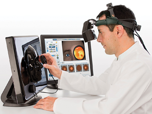
The Eyesi simulator from VRmagic (Mannheim, Germany) is a virtual reality system. When using the surgical modules, students operate on a sensor-laden “eye” while looking through a viewer resembling a microscope that provides a virtual picture of the simulated surgery they’re performing. One system is designed to simulate posterior segment surgeries, including a recently added retinal detachment surgery module; another system recreates anterior segment surgeries, especially cataract surgery. Recently, the company has released an indirect ophthalmoscope simulator, designed to give students practice in evaluation and diagnosis of disease.
Markus Schill, CEO at VRmagic, says that one of the company’s most important recent developments has been the inclusion of courseware with the systems. “At first, people who purchased the systems weren’t sure how to use them most effectively,” he says. “Now when you buy one of our simulators, it comes with a complete curriculum that starts your training at the most basic level, gradually adding skills and increasing the difficulty level.” He adds that a recent study in England found that students who trained on the simulator had significantly fewer complications in the OR.
Ophthalmoscopy
Mr. Schill says the new indirect ophthalmoscope simulator uses even more advanced technology than the surgical systems. “The indirect ophthalmoscope is an augmented reality system that mixes live video with digital images,” he explains. “The user wears a device mounted on an indirect ophthalmoscope headband. It feels pretty much like wearing the real thing, but the user is actually looking at video images captured by a camera in the frame. So, if you look around the room, you see the room. But when you turn your head toward the patient mask in front of you, the head-mounted display picks up and tracks digital markers on the mask, painting the digital patient into the video images. Similarly, when you bring your indirect lens into the optical path, the device paints the image of the retina that you would see onto the lens, matching your movements and angling of the lens. [The software also allows the user to choose lenses of different magnifications.] The feedback we’ve received so far is that it’s incredibly realistic.” (To see a video of the system in action, visit
vrmagic.com/simulators/eyesi-indirect.)
“In this system, the first tier teaches device handling, addressing issues such as: How do I hold the lens correctly? The second tier teaches what a normal eye looks like, and variations of that. The third tier teaches the user signs of basic pathologies and optic disc classification. The fourth tier simulates working in a hospital. You’re given a short patient history and do an exam; then you answer multiple choice questions. We’re also developing modules that will train users to detect and identify tumors and other pathologies. Currently there are 100 cases in the system, so you can see 100 digital patients before seeing real patients.”
Regarding the alternatives to virtual reality, Mr. Schill says all practice methods are useful and valid. “The major advantage of a system like ours is that there’s a curriculum embedded, so you’re not just practicing,” he says. “The system monitors everything you do, even factors such as how hard you’re pressing on the tissue, and evaluates your work. It provides hints and tips, telling the user what he or she did wrong. It’s a more complete learning environment, meant to take some of the training load off of the live teacher.”
Going Analog
|
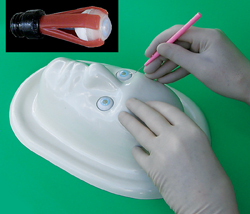
A completely different approach to surgical practice is epitomized by the Phak-i Surgical Practice Eye, made by Eye Care and Cure USA, in Tucson, Ariz. The Phak-i (pronounced “fake eye”) system includes the I-BORG, a life-sized face form, and Phak-i practice eyes that fit in the face form. The Phak-i is dimensionally and anatomically correct, and made of materials that when cut or manipulated closely approximate the feel of actually surgery on a human eye.
“I-BORG stands for Individual Basic Ophthalmic Research Guy (or Gal),” explains Johan Van Dalen, MD, PhD, CEO of Eye Care and Cure. “The reusable face form has a drain and a retaining basin around the face, so when there’s fluid coming from your phaco, it drains through a hole and vinyl tubing into a bag.”
Dr. Van Dalen says the cornea is designed to mimic a human cornea. “It’s half a millimeter thick in the central portion and 0.8 mm thick in the periphery,” he explains. “Of course, you can do multiple corneal procedures on one Phak-i. You can do six or seven incisions and practice suturing them; make scleral flaps and scleral tunnels; practice intravitreal injections; repair corneal or scleral lacerations; and perform limbal relaxing incisions. You can even do corneal transplants. Doctors frequently tell us that the cornea feels very real.
“The Phak-i has a lens with an anterior and posterior capsule and a dilated 8-mm pupil so you can do a capsulotomy,” he continues. “There’s no fluid inside the regular Phak-i, but if you want to do a cataract procedure you can inject fluid with a needle and it won’t leak. Of course, you can only do one capsulotomy and phacoemulsification of each lens, but you can repeat lens insertion and removal. You can also practice using capsular tension rings and iris expanders and hooks.”
Different Models, Different Skills
Dr. Van Dalen notes that the company produces multiple models. “One model has four muscles attached to it so you can do resection,” he says. “Another model has three retinal layers so you can practice retinal peeling. We also have a set of six Phak-i’s with different axial lengths. And because devices can now be inserted into the trabecular meshwork to improve aqueous flow, we’ve developed a trabecular meshwork equivalent that will be available this spring.” Other models designed to allow practice of specific procedures such as DSEK are also under development.
Dr. Van Dalen sees multiple advantages to using a simple and inexpensive model for surgical practice. “Animal eyes can be problematic, and virtual systems are much more expensive, limiting their availability,” he notes. “And with our Phak-i you can use your own instruments—your own phaco system, needles, forceps and so forth. You can’t do that in a virtual system. Our eyes also duplicate the feel of real surgery, and residents can take them home to practice on.”
The cost of one Phak-i is about $15; they come in two-packs and four-packs. The I-BORG face form is $135 and comes with the fluid retention bag and vinyl tubing. All components are made in the United States from non-toxic materials, and the eyes are fully recyclable. REVIEW


