Diabetic macular edema and proliferative diabetic retinopathy are a significant burden on the health care system, in part because they’re usually diagnosed and treated after significant damage has already been done. Now, an ophthalmologist has come up with an approach that may allow internists to inexpensively identify diabetic patients with early diabetic macular edema and proliferative diabetic retinopathy, prior to noticeable impact on vision, so that treatment can be started even before symptoms become evident.
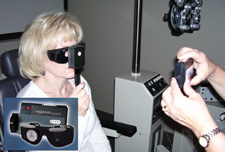
Shantan Reddy, MD, MPH, who practices at the DuPage Medical Group in Chicago, realized that an inexpensive test sometimes used to detect early macular degeneration could also detect early DME in diabetic patients. The test involves stressing the macula with bright light and then timing the visual recovery. Dr. Reddy uses two instruments to accomplish this: the Retinal Acuity Meter and the Brightness Acuity Meter, both developed and manufactured by AMA Optics in Miami. (Dr. Reddy has no financial interest in the company or products.)
“Diabetic retinopathy and diabetic macular edema are vascular problems, but there are also very subtle neurogenic or neurodegenerative problems in the retina,” says Dr. Reddy. “The problem with diabetic macular edema and diabetes in general is that we catch it very late. By the time these patients come to see us or are referred to us, they’ve already lost moderate to significant vision. So our goal should be to try to find these patients before they have vision loss.
“One of the ways we can do this is to use the photo-stress test,” he continues. “Stressing the macula with a bright light and measuring how long it takes vision to recover has been used for years to evaluate macular degeneration. In fact, this strategy reveals many kinds of macular pathology.”
How the Test Works
Dr. Reddy says the test is accomplished using the Retinal Acuity Meter, Brightness Acuity Meter and a stopwatch. “The patient’s best retinal acuity is measured with the Retinal Acuity Meter,” he says. “Then one eye is exposed to bright light using the Brightness Acuity Meter set on high for 30 seconds, after which the patient’s vision is re-tested with the RAM. The time required for the patient to return to the best retinal acuity level is the recovery time. Patients with early damage pathology simply take a few seconds longer to recover vision.
“Best of all, because of the way the test works, it’s unaffected by the presence of cataract, which might otherwise confound the results,” he says. “That’s where the RAM comes in. It uses an acuity measurement process involving the patient reading through uniform pinholes; the letters in the reading portion of the test are black Snellen or ETDRS letters against an illuminated background. The RAM acts like a potential acuity meter, but it’s easier to use and is more accurate in eyes with co-morbid disease. In fact, it’s being used right now by many cataract surgeons looking at visual potential to decide whether a cataract should come out. We’re just combining that with the Brightness Acuity Meter to photostress the macula in patients who may have diabetic macular edema.”
Dr. Reddy and colleague Kevin Chen, MD, conducted a pilot study to see whether this approach was clinically viable. They compared the results of a 30-second photostress test on two age-matched groups; the members of one group were those diagnosed with diabetic macular edema using OCT and ophthalmoscopy. The study involved 143 eyes of 84 patients, mean age 58.2 years (range 24 to 86), of which 48 eyes (33.6 percent) were found to have macular edema. There was no difference in retinal acuity between the DME and healthy groups, indicating that the DME patients had not suffered detectable functional loss at the time of testing.
The data showed that healthy eyes had a mean recovery time of 35.3 seconds (median: 32 seconds); eyes with diabetic macular edema had a mean photostress recovery time of 38.9 seconds (median: 34 seconds). The difference was statistically significant (p=0.04). Other factors, such as BCVA, central foveal thickness and retinal acuity did not correlate with recovery time.
Dr. Reddy notes that although the difference in recovery times is only a couple of seconds, they were able to detect the difference to statistical significance in the study. “You wouldn’t expect the difference to be huge,” he says. “After all, we’re identifying people who have very early macular edema. The difference would be much larger if they had more pathology, but once they have a lot of pathology, they’ll be coming in anyway. This will help us find the ones who need treatment but don’t know it yet.”
Dr. Reddy says that because this pilot study involved a limited number of eyes, the numbers didn’t allow them to determine the sensitivity or specificity of the test. “That wasn’t the intent of our small study,” he says. “We just wanted to see if there was sufficient statistical correlation to justify further research. But our data clearly shows that this test may provide warning when an exam of a diabetic patient would otherwise show nothing wrong. In our opinion, that makes it a potentially very valuable tool.”
Getting the Jump on DME
“Ultimately, we want internists to use this,” says Dr. Reddy. “Internists don’t usually check vision or look inside the eye to see if there’s any sign of macular edema. But this test is easy to perform, and if there’s a delayed photostress recovery time, they can refer the patient to an ophthalmologist. A non-mydriatic fundus camera could serve this purpose as well, but those are expensive and bulky, and technicians have to be trained to use them. They’re not really feasible for internists to have in the office, and ultimately, internists are the front lines in our battle against diabetic retinopathy.”
|
“This test should be easy for internists to use because they can train their technicians to do it—or even volunteers. It’s a very easy, non-invasive test, and the patients don’t need to be in an exam room. A patient could fill out his registration and then do the screening right then and there. The room doesn’t even have to be dark. The patient puts on the [pinhole] glasses and you conduct the test. That’s it. Furthermore, the equipment is relatively inexpensive—about $1,000 for the system.”
Dr. Reddy doesn’t believe patients will become overconfident about their prognosis if they pass the test. “Part of the internists’ job is to emphasize the importance of yearly dilated exams for diabetic patients, even if they pass the test,” he says. “In the meantime, internists will be able to go beyond simply asking whether the patient has any vision problems. They’ll be able to capture many patients who have no apparent vision problems but do have macular edema that needs treatment. Those are the most vulnerable patients, and the ones with the most visual potential. Those are the ones who stand to gain the most from treatment.” REVIEW


